8U7C | pdb_00008u7c

Engineered NEMO minimal IKK-binding domain


Experimental Data Snapshot

  • Method: X-RAY DIFFRACTION
  • Resolution: 1.44 Å
  • R-Value Free: 
    0.202 (Depositor), 0.203 (DCC) 
  • R-Value Work: 
    0.173 (Depositor), 0.173 (DCC) 
  • R-Value Observed: 
    0.175 (Depositor) 

wwPDB Validation   3D Report Full Report


This is version 1.1 of the entry. See complete history


Literature

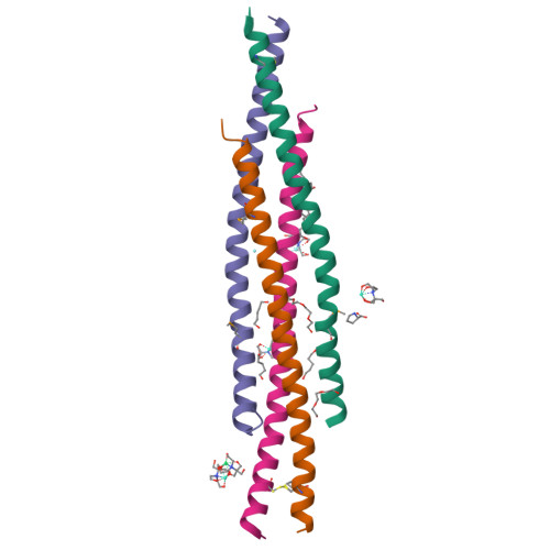
The structure of a NEMO construct engineered for screening reveals novel determinants of inhibition.

Kennedy, A.E.Barczewski, A.H.Arnoldy, C.R.Pennington, J.P.Tiernan, K.A.Hidalgo, M.B.Reilly, C.C.Wongsri, T.Ragusa, M.J.Grigoryan, G.Mierke, D.F.Pellegrini, M.

(2025) Structure 

  • DOI: https://doi.org/10.1016/j.str.2025.01.010
  • Primary Citation of Related Structures:  
    8U7C

  • PubMed Abstract: 

    NEMO is an essential component in the activation of the canonical nuclear factor κB (NF-κB) pathway and exerts its function by recruiting the IκB kinases (IKK) to the IKK complex. Inhibition of the NEMO/IKKs interaction is an attractive therapeutic paradigm for diseases related to NF-κB mis-regulation, but a difficult endeavor because of the extensive protein-protein interface. Here we report the design and characterization of novel engineered constructs of the IKK-binding domain of NEMO, programmed to render this difficult protein domain amenable to NMR measurements and crystallization, while preserving its biological function. ZipNEMO binds IKKβ with nanomolar affinity, is amenable to heteronuclear nuclear magnetic resonance (NMR) techniques and structure determination by X-ray crystallography. We show that NMR spectra of zipNEMO allow to detect inhibitor binding in solution and resonance assignment. The crystal structure of zipNEMO reveals a novel ligand binding motif and the adaptability of the binding pocket and inspired the design of new peptide inhibitors.


  • Organizational Affiliation

    Department of Chemistry, Dartmouth College, Hanover, NH 03755, USA.


Macromolecules
Find similar proteins by:  (by identity cutoff)  |  3D Structure
Entity ID: 1
MoleculeChains Sequence LengthOrganismDetailsImage
Engineered NEMO minimal IKK-binding domain
A, B, C, D
67Homo sapiensMutation(s): 0 
UniProt & NIH Common Fund Data Resources
Find proteins for Q9Y6K9 (Homo sapiens)
Explore Q9Y6K9 
Go to UniProtKB:  Q9Y6K9
PHAROS:  Q9Y6K9
GTEx:  ENSG00000269335 
Entity Groups  
Sequence Clusters30% Identity50% Identity70% Identity90% Identity95% Identity100% Identity
UniProt GroupQ9Y6K9
Sequence Annotations
Expand
  • Reference Sequence
Small Molecules
Ligands 7 Unique
IDChains Name / Formula / InChI Key2D Diagram3D Interactions
BTB
Query on BTB

Download Ideal Coordinates CCD File 
H [auth A],
U [auth D],
V [auth D],
W [auth D],
X [auth D]
2-[BIS-(2-HYDROXY-ETHYL)-AMINO]-2-HYDROXYMETHYL-PROPANE-1,3-DIOL
C8 H19 N O5
OWMVSZAMULFTJU-UHFFFAOYSA-N
PG4
Query on PG4

Download Ideal Coordinates CCD File 
L [auth B],
T [auth D]
TETRAETHYLENE GLYCOL
C8 H18 O5
UWHCKJMYHZGTIT-UHFFFAOYSA-N
ER3
Query on ER3

Download Ideal Coordinates CCD File 
Q [auth D]ERBIUM (III) ION
Er
JHFPQYFEJICGKC-UHFFFAOYSA-N
TB
Query on TB

Download Ideal Coordinates CCD File 
E [auth A],
O [auth D],
P [auth D]
TERBIUM(III) ION
Tb
HKCRVXUAKWXBLE-UHFFFAOYSA-N
PRO
Query on PRO

Download Ideal Coordinates CCD File 
I [auth A],
N [auth B]
PROLINE
C5 H9 N O2
ONIBWKKTOPOVIA-BYPYZUCNSA-N
9JE
Query on 9JE

Download Ideal Coordinates CCD File 
F [auth A],
G [auth A],
K [auth B],
M [auth B],
S [auth D]
pentane-1,5-diol
C5 H12 O2
ALQSHHUCVQOPAS-UHFFFAOYSA-N
YT3
Query on YT3

Download Ideal Coordinates CCD File 
J [auth B],
R [auth D]
YTTRIUM (III) ION
Y
GRTBAGCGDOYUBE-UHFFFAOYSA-N
Modified Residues  1 Unique
IDChains TypeFormula2D DiagramParent
MSE
Query on MSE
A, B, C, D
L-PEPTIDE LINKINGC5 H11 N O2 SeMET
Experimental Data & Validation

Experimental Data

  • Method: X-RAY DIFFRACTION
  • Resolution: 1.44 Å
  • R-Value Free:  0.202 (Depositor), 0.203 (DCC) 
  • R-Value Work:  0.173 (Depositor), 0.173 (DCC) 
  • R-Value Observed: 0.175 (Depositor) 
Space Group: P 1
Unit Cell:
Length ( Å )Angle ( ˚ )
a = 37.514α = 92.62
b = 40.892β = 106.14
c = 50.15γ = 98.87
Software Package:
Software NamePurpose
PHENIXrefinement
Cootmodel building
autoPROCdata reduction
STARANISOdata scaling
PHENIXphasing

Structure Validation

View Full Validation Report


Entry History & Funding Information

Deposition Data


Funding OrganizationLocationGrant Number
National Institutes of Health/National Institute Of Allergy and Infectious Diseases (NIH/NIAID)United States1R01GM133844-01

Revision History  (Full details and data files)

  • Version 1.0: 2025-01-22
    Type: Initial release
  • Version 1.1: 2025-02-19
    Changes: Database references