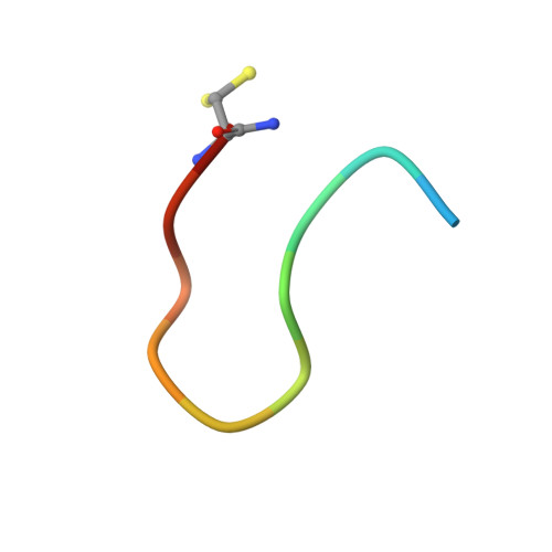
8PKX

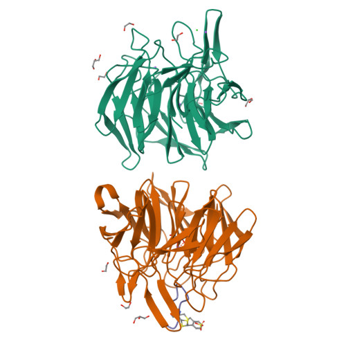
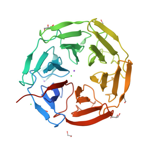
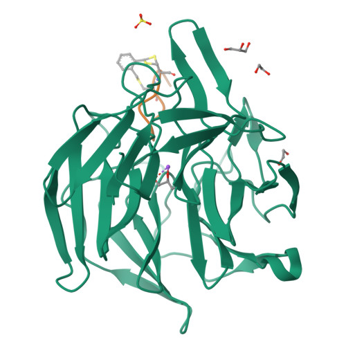
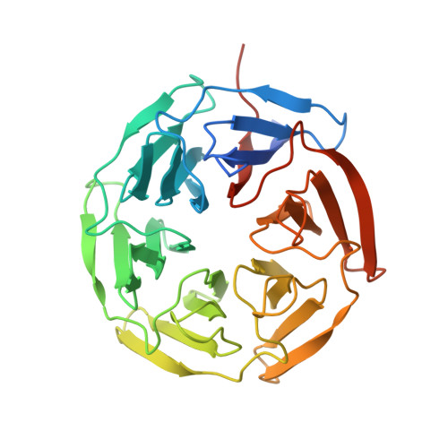
Kelch domain of KEAP1 in complex with a ortho-dimethylbenzene linked cyclic peptide 11 (ortho-WRCNPETaEC).


Experimental Data Snapshot

  • Method: X-RAY DIFFRACTION
  • Resolution: 1.79 Å
  • R-Value Free: 0.210 
  • R-Value Work: 0.175 
  • R-Value Observed: 0.176 

Starting Model: experimental
View more details

wwPDB Validation   3D Report Full Report


Ligand Structure Quality Assessment 


This is version 1.3 of the entry. See complete history


Literature

Computational Prediction of Cyclic Peptide Structural Ensembles and Application to the Design of Keap1 Binders.

Fonseca Lopez, F.Miao, J.Damjanovic, J.Bischof, L.Braun, M.B.Ling, Y.Hartmann, M.D.Lin, Y.S.Kritzer, J.A.

(2023) J Chem Inf Model 63: 6925-6937

  • DOI: https://doi.org/10.1021/acs.jcim.3c01337
  • Primary Citation of Related Structures:  
    8PKU, 8PKV, 8PKW, 8PKX

  • PubMed Abstract: 

    The Nrf2 transcription factor is a master regulator of the cellular response to oxidative stress, and Keap1 is its primary negative regulator. Activating Nrf2 by inhibiting the Nrf2-Keap1 protein-protein interaction has shown promise for treating cancer and inflammatory diseases. A loop derived from Nrf2 has been shown to inhibit Keap1 selectively, especially when cyclized, but there are no reliable design methods for predicting an optimal macrocyclization strategy. In this work, we employed all-atom, explicit-solvent molecular dynamics simulations with enhanced sampling methods to predict the relative degree of preorganization for a series of peptides cyclized with a set of bis-thioether "staples". We then correlated these predictions to experimentally measured binding affinities for Keap1 and crystal structures of the cyclic peptides bound to Keap1. This work showcases a computational method for designing cyclic peptides by simulating and comparing their entire solution-phase ensembles, providing key insights into designing cyclic peptides as selective inhibitors of protein-protein interactions.


  • Organizational Affiliation

    Department of Chemistry, Tufts University, Medford, Massachusetts 02155, United States.


Macromolecules
Find similar proteins by:  (by identity cutoff)  |  3D Structure
Entity ID: 1
MoleculeChains Sequence LengthOrganismDetailsImage
Kelch-like ECH-associated protein 1
A, B
316Homo sapiensMutation(s): 0 
Gene Names: KEAP1INRF2KIAA0132KLHL19
UniProt & NIH Common Fund Data Resources
Find proteins for Q14145 (Homo sapiens)
Explore Q14145 
Go to UniProtKB:  Q14145
PHAROS:  Q14145
GTEx:  ENSG00000079999 
Entity Groups  
Sequence Clusters30% Identity50% Identity70% Identity90% Identity95% Identity100% Identity
UniProt GroupQ14145
Sequence Annotations
Expand
  • Reference Sequence

Find similar proteins by:  Sequence   |   3D Structure  

Entity ID: 2
MoleculeChains Sequence LengthOrganismDetailsImage
CP11C [auth P]12Homo sapiensMutation(s): 0 
Entity Groups  
Sequence Clusters30% Identity50% Identity70% Identity90% Identity95% Identity100% Identity
Sequence Annotations
Expand
  • Reference Sequence
Small Molecules
Ligands 6 Unique
IDChains Name / Formula / InChI Key2D Diagram3D Interactions
ZK2 (Subject of Investigation/LOI)
Query on ZK2

Download Ideal Coordinates CCD File 
U [auth P](2-methylphenyl)methanol
C8 H10 O
XPNGNIFUDRPBFJ-UHFFFAOYSA-N
SO4
Query on SO4

Download Ideal Coordinates CCD File 
O [auth B]SULFATE ION
O4 S
QAOWNCQODCNURD-UHFFFAOYSA-L
GOL
Query on GOL

Download Ideal Coordinates CCD File 
H [auth A],
K [auth A],
Q [auth B],
S [auth B]
GLYCEROL
C3 H8 O3
PEDCQBHIVMGVHV-UHFFFAOYSA-N
EDO
Query on EDO

Download Ideal Coordinates CCD File 
F [auth A]
G [auth A]
I [auth A]
J [auth A]
R [auth B]
F [auth A],
G [auth A],
I [auth A],
J [auth A],
R [auth B],
T [auth B]
1,2-ETHANEDIOL
C2 H6 O2
LYCAIKOWRPUZTN-UHFFFAOYSA-N
CL
Query on CL

Download Ideal Coordinates CCD File 
D [auth A],
E [auth A]
CHLORIDE ION
Cl
VEXZGXHMUGYJMC-UHFFFAOYSA-M
NA
Query on NA

Download Ideal Coordinates CCD File 
L [auth A],
M [auth A],
N [auth A],
P [auth B]
SODIUM ION
Na
FKNQFGJONOIPTF-UHFFFAOYSA-N
Experimental Data & Validation

Experimental Data

  • Method: X-RAY DIFFRACTION
  • Resolution: 1.79 Å
  • R-Value Free: 0.210 
  • R-Value Work: 0.175 
  • R-Value Observed: 0.176 
  • Space Group: C 1 2 1
Unit Cell:
Length ( Å )Angle ( ˚ )
a = 162.43α = 90
b = 68.877β = 117.676
c = 78.252γ = 90
Software Package:
Software NamePurpose
PHENIXrefinement
XDSdata reduction
Cootmodel building
PHASERphasing

Structure Validation

View Full Validation Report


Ligand Structure Quality Assessment 


Entry History & Funding Information

Deposition Data


Funding OrganizationLocationGrant Number
Not funded--

Revision History  (Full details and data files)

  • Version 1.0: 2023-11-15
    Type: Initial release
  • Version 1.1: 2023-11-22
    Changes: Database references
  • Version 1.2: 2023-12-13
    Changes: Structure summary
  • Version 1.3: 2024-10-09
    Changes: Structure summary