6R1O | pdb_00006r1o

Crystal structure of E. coli seryl-tRNA synthetase complexed to a seryl sulfamoyl adenosine derivative


Experimental Data Snapshot

  • Method: X-RAY DIFFRACTION
  • Resolution: 2.60 Å
  • R-Value Free: 
    0.215 (Depositor), 0.220 (DCC) 
  • R-Value Work: 
    0.182 (Depositor), 0.180 (DCC) 
  • R-Value Observed: 
    0.184 (Depositor) 

Starting Model: experimental
View more details

wwPDB Validation   3D Report Full Report


Ligand Structure Quality Assessment 

Created with Raphaël 2.3.0Worse 01 BetterLigand structure goodness of fit to experimental dataBest fitted JPEClick on this verticalbar to view details

This is version 1.2 of the entry. See complete history


Literature

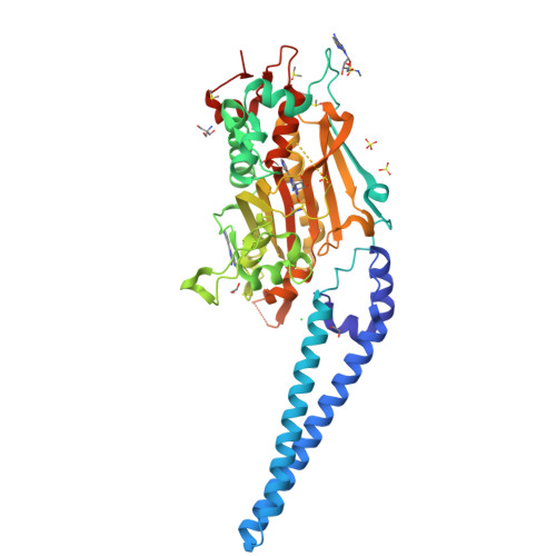
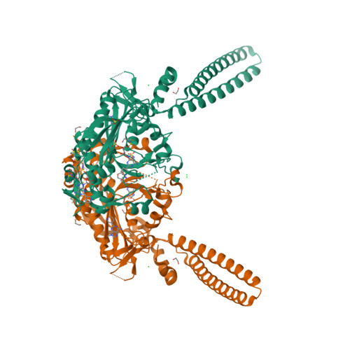
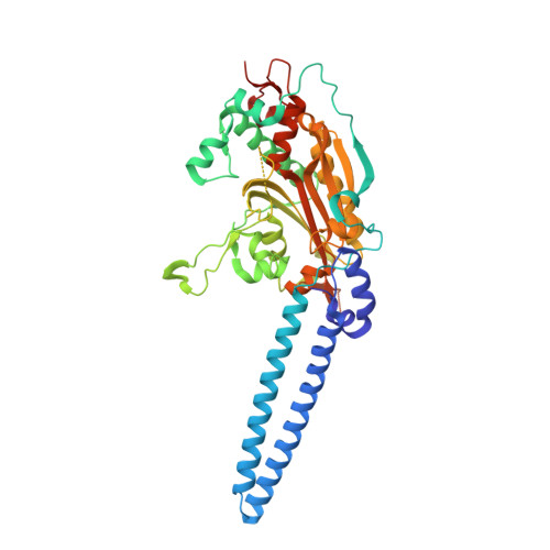
Structure-Guided Enhancement of Selectivity of Chemical Probe Inhibitors Targeting Bacterial Seryl-tRNA Synthetase.

Cain, R.Salimraj, R.Punekar, A.S.Bellini, D.Fishwick, C.W.G.Czaplewski, L.Scott, D.J.Harris, G.Dowson, C.G.Lloyd, A.J.Roper, D.I.

(2019) J Med Chem 62: 9703-9717

  • DOI: https://doi.org/10.1021/acs.jmedchem.9b01131
  • Primary Citation of Related Structures:  
    6R1M, 6R1N, 6R1O

  • PubMed Abstract: 

    Aminoacyl-tRNA synthetases are ubiquitous and essential enzymes for protein synthesis and also a variety of other metabolic processes, especially in bacterial species. Bacterial aminoacyl-tRNA synthetases represent attractive and validated targets for antimicrobial drug discovery if issues of prokaryotic versus eukaryotic selectivity and antibiotic resistance generation can be addressed. We have determined high-resolution X-ray crystal structures of the Escherichia coli and Staphylococcus aureus seryl-tRNA synthetases in complex with aminoacyl adenylate analogues and applied a structure-based drug discovery approach to explore and identify a series of small molecule inhibitors that selectively inhibit bacterial seryl-tRNA synthetases with greater than 2 orders of magnitude compared to their human homologue, demonstrating a route to the selective chemical inhibition of these bacterial targets.


  • Organizational Affiliation

    School of Life Sciences , University of Warwick , Gibbet Hill Road , Coventry CV4 7AL , United Kingdom.


Macromolecules
Find similar proteins by:  (by identity cutoff)  |  3D Structure
Entity ID: 1
MoleculeChains Sequence LengthOrganismDetailsImage
Serine--tRNA ligase437Escherichia coliMutation(s): 0 
Gene Names: serSECVG_02257
EC: 6.1.1.11
UniProt
Find proteins for P0A8L1 (Escherichia coli (strain K12))
Explore P0A8L1 
Go to UniProtKB:  P0A8L1
Entity Groups  
Sequence Clusters30% Identity50% Identity70% Identity90% Identity95% Identity100% Identity
UniProt GroupP0A8L1
Sequence Annotations
Expand
  • Reference Sequence
Small Molecules
Ligands 6 Unique
IDChains Name / Formula / InChI Key2D Diagram3D Interactions
JPE (Subject of Investigation/LOI)
Query on JPE

Download Ideal Coordinates CCD File 
B [auth A],
C [auth A],
D [auth A]
[(2~{R},3~{S},4~{R},5~{R})-5-(6-azanyl-2-pyridin-3-yl-purin-9-yl)-3,4-bis(oxidanyl)oxolan-2-yl]methyl ~{N}-[(2~{S})-2-azanyl-3-oxidanyl-propanoyl]sulfamate
C18 H22 N8 O8 S
AJLUWRJYNKUFKX-OBXKABKQSA-N
TRS
Query on TRS

Download Ideal Coordinates CCD File 
S [auth A]2-AMINO-2-HYDROXYMETHYL-PROPANE-1,3-DIOL
C4 H12 N O3
LENZDBCJOHFCAS-UHFFFAOYSA-O
SO4
Query on SO4

Download Ideal Coordinates CCD File 
G [auth A],
H [auth A],
I [auth A]
SULFATE ION
O4 S
QAOWNCQODCNURD-UHFFFAOYSA-L
DMS
Query on DMS

Download Ideal Coordinates CCD File 
J [auth A],
K [auth A],
L [auth A],
M [auth A]
DIMETHYL SULFOXIDE
C2 H6 O S
IAZDPXIOMUYVGZ-UHFFFAOYSA-N
EDO
Query on EDO

Download Ideal Coordinates CCD File 
N [auth A],
O [auth A],
P [auth A],
Q [auth A],
R [auth A]
1,2-ETHANEDIOL
C2 H6 O2
LYCAIKOWRPUZTN-UHFFFAOYSA-N
CL
Query on CL

Download Ideal Coordinates CCD File 
E [auth A],
F [auth A]
CHLORIDE ION
Cl
VEXZGXHMUGYJMC-UHFFFAOYSA-M
Binding Affinity Annotations 
IDSourceBinding Affinity
JPE BindingDB:  6R1O Kd: min: 290, max: 1920 (nM) from 2 assay(s)
IC50: 6650 (nM) from 1 assay(s)
Experimental Data & Validation

Experimental Data

  • Method: X-RAY DIFFRACTION
  • Resolution: 2.60 Å
  • R-Value Free:  0.215 (Depositor), 0.220 (DCC) 
  • R-Value Work:  0.182 (Depositor), 0.180 (DCC) 
  • R-Value Observed: 0.184 (Depositor) 
Space Group: P 61 2 2
Unit Cell:
Length ( Å )Angle ( ˚ )
a = 85.41α = 90
b = 85.41β = 90
c = 317.716γ = 120
Software Package:
Software NamePurpose
BUSTERrefinement
XDSdata reduction
autoPROCdata scaling
PHASERphasing

Structure Validation

View Full Validation Report


Ligand Structure Quality Assessment 

Created with Raphaël 2.3.0Worse 01 BetterLigand structure goodness of fit to experimental dataBest fitted JPEClick on this verticalbar to view details

Entry History & Funding Information

Deposition Data


Funding OrganizationLocationGrant Number
Medical Research Council (United Kingdom)United KingdomMR/M017893/1

Revision History  (Full details and data files)

  • Version 1.0: 2020-01-22
    Type: Initial release
  • Version 1.1: 2021-02-10
    Changes: Refinement description
  • Version 1.2: 2024-01-24
    Changes: Data collection, Database references, Refinement description