6H8J

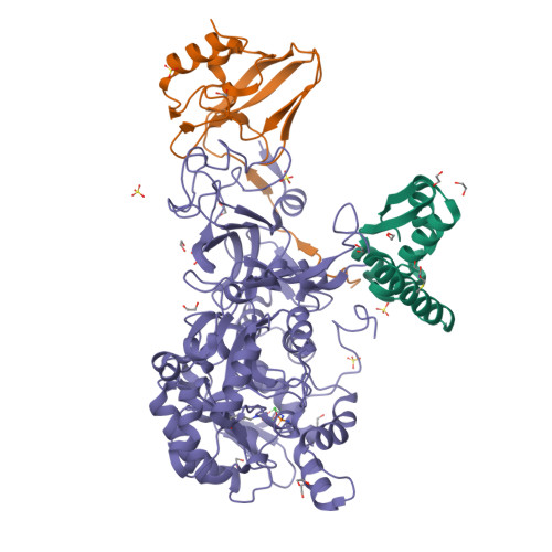
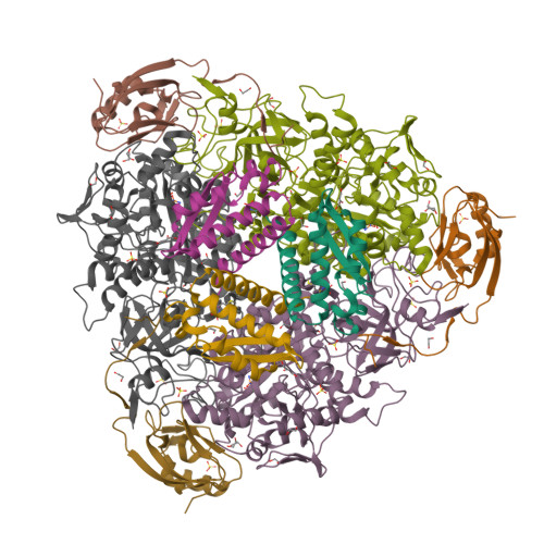
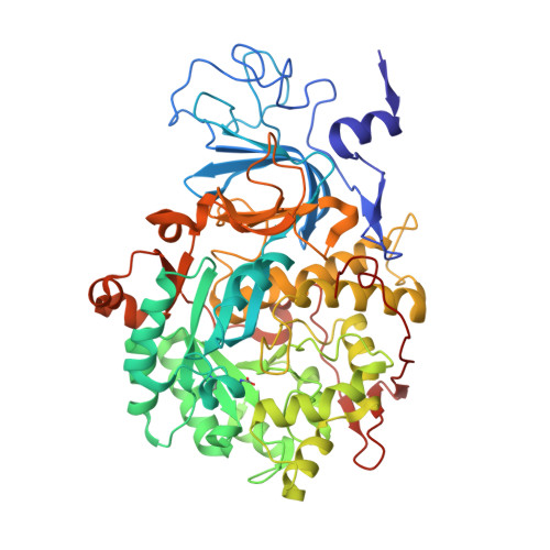
1.45 A resolution of Sporosarcina pasteurii urease inhibited in the presence of NBPTO


Experimental Data Snapshot

  • Method: X-RAY DIFFRACTION
  • Resolution: 1.45 Å
  • R-Value Free: 0.143 
  • R-Value Work: 0.124 
  • R-Value Observed: 0.125 

Starting Model: experimental
View more details

wwPDB Validation   3D Report Full Report


This is version 1.1 of the entry. See complete history


Literature

Insights into Urease Inhibition by N-( n-Butyl) Phosphoric Triamide through an Integrated Structural and Kinetic Approach.

Mazzei, L.Cianci, M.Contaldo, U.Ciurli, S.

(2019) J Agric Food Chem 67: 2127-2138

  • DOI: https://doi.org/10.1021/acs.jafc.8b04791
  • Primary Citation of Related Structures:  
    6H8J

  • PubMed Abstract: 

    The nickel-dependent enzyme urease represents a negative element for the efficiency of soil nitrogen fertilization as well as a virulence factor for a large number of pathogenic and antibiotic-resistant bacteria. The development of ever more efficient urease inhibitors demands knowledge of their modes of action at the molecular level. N-( n-Butyl)-phosphoric triamide (NBPTO) is the oxo-derivative of N-( n-butyl)-thiophosphoric triamide (NBPT), which is extensively employed in agriculture to increase the efficiency of urea-based fertilizers. The 1.45 Å resolution structure of the enzyme-inhibitor complex obtained upon incubation of Sporosarcina pasteurii urease (SPU) with NBPTO shows the presence of diamido phosphoric acid (DAP), generated upon enzymatic hydrolysis of NBPTO with the release of n-butyl amine. DAP is bound in a tridentate binding mode to the two Ni(II) ions in the active site of urease via two O atoms and an amide NH 2 group, whereas the second amide group of DAP points away from the metal center into the active-site channel. The mobile flap modulating the size of the active-site cavity is found in a disordered closed-open conformation. A kinetic characterization of the NBPTO-based inhibition of both bacterial (SPU) and plant ( Canavalia ensiformis or jack bean, JBU) ureases, carried out by calorimetric measurements, indicates the occurrence of a reversible slow-inhibition mode of action. The latter is characterized by a very small value of the equilibrium dissociation constant of the urease-DAP complex caused, in turn, by the large rate constant for the formation of the enzyme-inhibitor complex. The much greater capability of NBPTO to inhibit urease, as compared with that of NBPT, is thus not caused by the presence of a P═O moiety versus a P═S moiety, as previously suggested, but rather by the readiness of NBPTO to react with urease without the need to convert one of the P-NH 2 amide moieties to its P-OH acid derivative, as in the case of NBPT. The latter process is indeed characterized by a very small equilibrium constant that reduces drastically the concentration of the active form of the inhibitor in the case of NBPT. This indicates that high-efficiency phosphoramide-based urease inhibitors must have at least one O atom bound to the central P atom in order for the molecule to efficiently and rapidly bind to the dinickel center of the enzyme.


  • Organizational Affiliation

    Laboratory of Bioinorganic Chemistry, Department of Pharmacy and Biotechnology , University of Bologna , 40126 Bologna , Italy.


Macromolecules
Find similar proteins by:  (by identity cutoff)  |  3D Structure
Entity ID: 1
MoleculeChains Sequence LengthOrganismDetailsImage
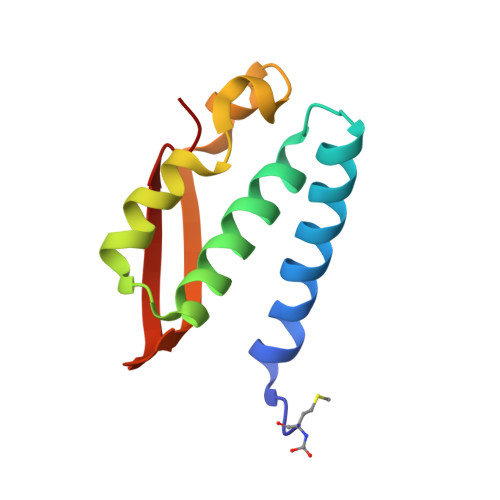
Urease subunit gamma100Sporosarcina pasteuriiMutation(s): 0 
EC: 3.5.1.5
UniProt
Find proteins for P41022 (Sporosarcina pasteurii)
Explore P41022 
Go to UniProtKB:  P41022
Entity Groups  
Sequence Clusters30% Identity50% Identity70% Identity90% Identity95% Identity100% Identity
UniProt GroupP41022
Sequence Annotations
Expand
  • Reference Sequence
Find similar proteins by:  (by identity cutoff)  |  3D Structure
Entity ID: 2
MoleculeChains Sequence LengthOrganismDetailsImage
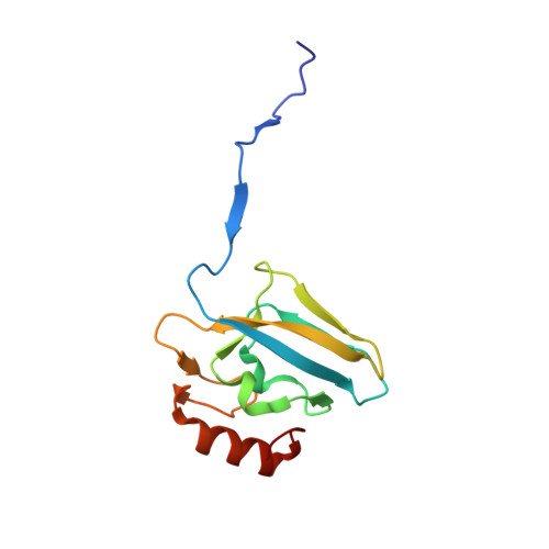
Urease subunit beta122Sporosarcina pasteuriiMutation(s): 0 
EC: 3.5.1.5
UniProt
Find proteins for P41021 (Sporosarcina pasteurii)
Explore P41021 
Go to UniProtKB:  P41021
Entity Groups  
Sequence Clusters30% Identity50% Identity70% Identity90% Identity95% Identity100% Identity
UniProt GroupP41021
Sequence Annotations
Expand
  • Reference Sequence
Find similar proteins by:  (by identity cutoff)  |  3D Structure
Entity ID: 3
MoleculeChains Sequence LengthOrganismDetailsImage
Urease subunit alpha570Sporosarcina pasteuriiMutation(s): 0 
EC: 3.5.1.5
UniProt
Find proteins for P41020 (Sporosarcina pasteurii)
Explore P41020 
Go to UniProtKB:  P41020
Entity Groups  
Sequence Clusters30% Identity50% Identity70% Identity90% Identity95% Identity100% Identity
UniProt GroupP41020
Sequence Annotations
Expand
  • Reference Sequence
Small Molecules
Ligands 4 Unique
IDChains Name / Formula / InChI Key2D Diagram3D Interactions
SO4
Query on SO4

Download Ideal Coordinates CCD File 
H [auth A],
J [auth B],
K [auth B],
V [auth C],
W [auth C]
SULFATE ION
O4 S
QAOWNCQODCNURD-UHFFFAOYSA-L
2PA
Query on 2PA

Download Ideal Coordinates CCD File 
X [auth C]DIAMIDOPHOSPHATE
H5 N2 O2 P
ANCLJVISBRWUTR-UHFFFAOYSA-N
EDO
Query on EDO

Download Ideal Coordinates CCD File 
D [auth A]
E [auth A]
F [auth A]
G [auth A]
I [auth B]
D [auth A],
E [auth A],
F [auth A],
G [auth A],
I [auth B],
N [auth C],
O [auth C],
P [auth C],
Q [auth C],
R [auth C],
S [auth C],
T [auth C],
U [auth C]
1,2-ETHANEDIOL
C2 H6 O2
LYCAIKOWRPUZTN-UHFFFAOYSA-N
NI
Query on NI

Download Ideal Coordinates CCD File 
L [auth C],
M [auth C]
NICKEL (II) ION
Ni
VEQPNABPJHWNSG-UHFFFAOYSA-N
Modified Residues  2 Unique
IDChains TypeFormula2D DiagramParent
CXM
Query on CXM
A
L-PEPTIDE LINKINGC6 H11 N O4 SMET
KCX
Query on KCX
C
L-PEPTIDE LINKINGC7 H14 N2 O4LYS
Experimental Data & Validation

Experimental Data

  • Method: X-RAY DIFFRACTION
  • Resolution: 1.45 Å
  • R-Value Free: 0.143 
  • R-Value Work: 0.124 
  • R-Value Observed: 0.125 
  • Space Group: P 63 2 2
Unit Cell:
Length ( Å )Angle ( ˚ )
a = 131.446α = 90
b = 131.446β = 90
c = 188.716γ = 120
Software Package:
Software NamePurpose
REFMACrefinement
XDSdata reduction
Aimlessdata scaling
REFMACphasing

Structure Validation

View Full Validation Report


Entry History 

Deposition Data

Revision History  (Full details and data files)

  • Version 1.0: 2019-06-12
    Type: Initial release
  • Version 1.1: 2024-01-17
    Changes: Data collection, Database references, Derived calculations, Refinement description