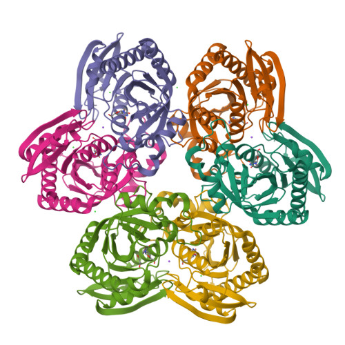
X-ray structure of uridine phosphorylase from Vibrio cholerae in complex with uridine at 1.03 A resolution
Prokofev, I.I., Lashkov, A.A., Gabdulkhakov, A.G., Dontsova, M.V., Betzel, C., Mikhailov, A.M.To be published.
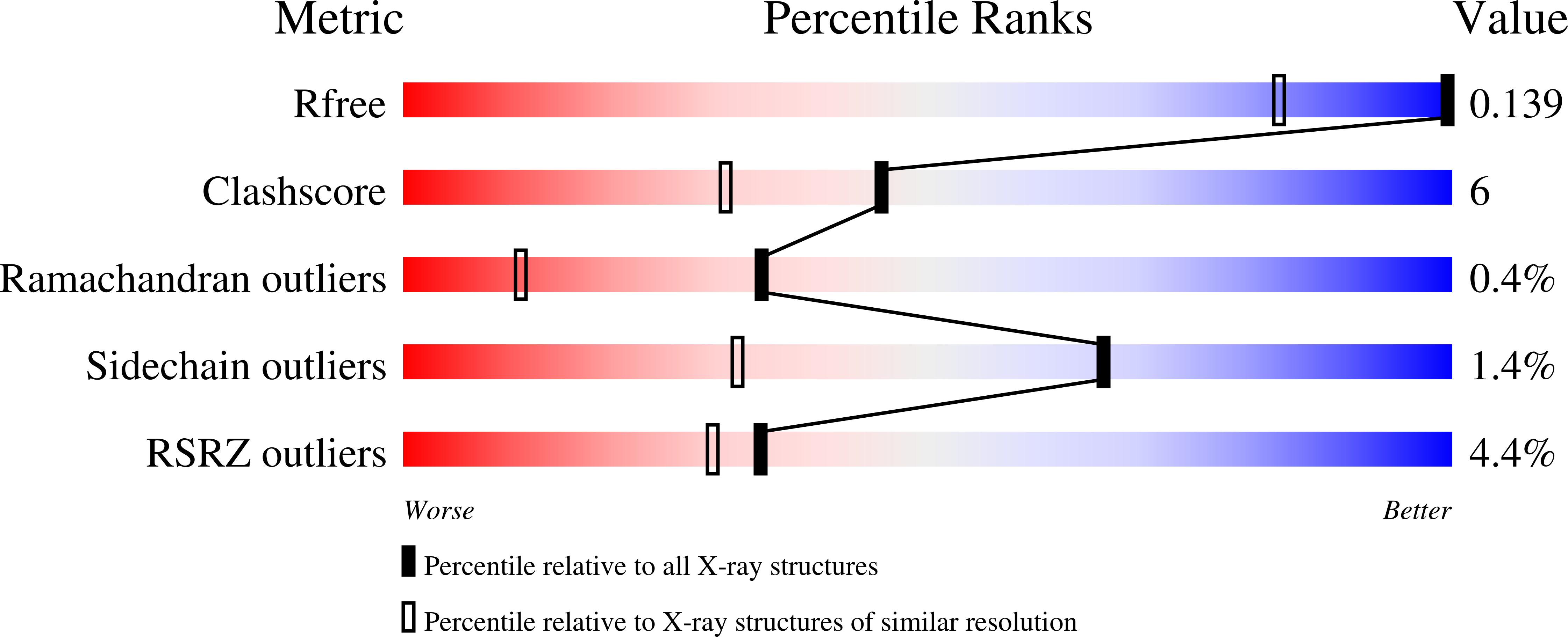
Experimental Data Snapshot
Starting Model: experimental
View more details
Entity ID: 1 | |||||
|---|---|---|---|---|---|
| Molecule | Chains | Sequence Length | Organism | Details | Image |
| Uridine phosphorylase | 253 | Vibrio cholerae | Mutation(s): 0 Gene Names: udp, udp_1, B2J67_05455, B2J68_05210, B2J71_05165, BFX32_04820, BTY66_05555, CEF09_03900, DN30_1909, EN12_05055... EC: 2.4.2.3 | 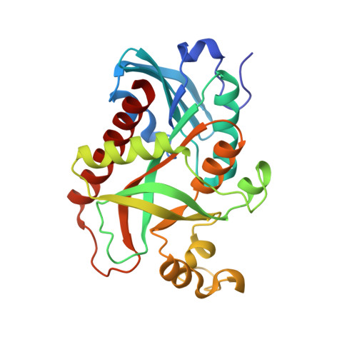 | |
UniProt | |||||
Find proteins for Q9K4U1 (Vibrio cholerae) Explore Q9K4U1 Go to UniProtKB: Q9K4U1 | |||||
Entity Groups | |||||
| Sequence Clusters | 30% Identity50% Identity70% Identity90% Identity95% Identity100% Identity | ||||
| UniProt Group | Q9K4U1 | ||||
Sequence AnnotationsExpand | |||||
| |||||
| Ligands 5 Unique | |||||
|---|---|---|---|---|---|
| ID | Chains | Name / Formula / InChI Key | 2D Diagram | 3D Interactions | |
| URI Query on URI | G [auth A] I [auth B] K [auth C] O [auth D] S [auth E] | URIDINE C9 H12 N2 O6 DRTQHJPVMGBUCF-XVFCMESISA-N |  | ||
| EDO Query on EDO | U [auth E] | 1,2-ETHANEDIOL C2 H6 O2 LYCAIKOWRPUZTN-UHFFFAOYSA-N |  | ||
| CL Query on CL | J [auth B] L [auth C] M [auth C] P [auth D] Q [auth D] | CHLORIDE ION Cl VEXZGXHMUGYJMC-UHFFFAOYSA-M |  | ||
| MG Query on MG | R [auth D] | MAGNESIUM ION Mg JLVVSXFLKOJNIY-UHFFFAOYSA-N |  | ||
| NA Query on NA | H [auth A], N [auth C], T [auth E] | SODIUM ION Na FKNQFGJONOIPTF-UHFFFAOYSA-N |  | ||
| Length ( Å ) | Angle ( ˚ ) |
|---|---|
| a = 64.323 | α = 110.56 |
| b = 72.039 | β = 107.53 |
| c = 89.193 | γ = 85.83 |
| Software Name | Purpose |
|---|---|
| PHENIX | refinement |
| XSCALE | data scaling |
| PDB_EXTRACT | data extraction |
| MOLREP | phasing |
| XSCALE | data reduction |
| Funding Organization | Location | Grant Number |
|---|---|---|
| RFBR | Russian Federation | 14-04-00952a |
| Grant of President of Russian Federation | Russian Federation | MK-9246.2016.3 |