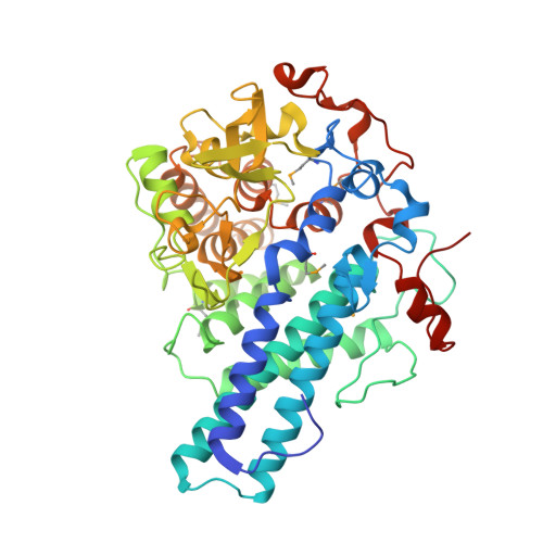
5CX8

Structure of RagB, a major immunodominant virulence factor of Porphyromonas gingivalis.


Experimental Data Snapshot

  • Method: X-RAY DIFFRACTION
  • Resolution: 2.40 Å
  • R-Value Free: 0.208 
  • R-Value Work: 0.178 
  • R-Value Observed: 0.178 

wwPDB Validation   3D Report Full Report


Ligand Structure Quality Assessment 


This is version 2.1 of the entry. See complete history


Literature

Structure of RagB, a major immunodominant outer-membrane surface receptor antigen of Porphyromonas gingivalis.

Goulas, T.Garcia-Ferrer, I.Hutcherson, J.A.Potempa, B.A.Potempa, J.Scott, D.A.Xavier Gomis-Ruth, F.

(2016) Mol Oral Microbiol 31: 472-485

  • DOI: https://doi.org/10.1111/omi.12140
  • Primary Citation of Related Structures:  
    5CX8

  • PubMed Abstract: 

    Porphyromonas gingivalis is the main causative agent of periodontitis. It deregulates the inflammatory and innate host immune responses through virulence factors, which include the immunodominant outer-membrane surface receptor antigens A (PgRagA) and B (PgRagB), co-transcribed from the rag pathogenicity island. The former is predicted to be a Ton-dependent porin-type translocator but the targets of this translocation and the molecular function of PgRagB are unknown. Phenomenologically, PgRagB has been linked with epithelial cell invasion and virulence according to murine models. It also acts as a Toll-like receptor agonist and promotes multiple mediators of inflammation. Hence, PgRagB is a candidate for the development of a periodontitis vaccine, which would be facilitated by the knowledge of its atomic structure. Here, we crystallized and solved the structure of 54-kDa PgRagB, which revealed a single domain centered on a curved helical scaffold. It consists of four tetratrico peptide repeats (TPR1-4), each arranged as two helices connected by a linker, plus two extra downstream capping helices. The concave surface bears four large intertwined irregular inserts (A-D), which contribute to an overall compact moiety. Overall, PgRagB shows substantial structural similarity with Bacteroides thetaiotaomicron SusD and Tannerella forsythia NanU, which are, respectively, engaged in binding and uptake of malto-oligosaccharide/starch and sialic acid. This suggests a similar sugar-binding function for PgRagB for uptake by the cognate PgRagA translocator, and, consistently, three potential monosaccharide-binding sites were tentatively assigned on the molecular surface.


  • Organizational Affiliation

    Proteolysis Laboratory, Department of Structural Biology ('María de Maeztu' Unit of Excellence), Molecular Biology Institute of Barcelona, CSIC, Barcelona, Spain.


Macromolecules
Find similar proteins by:  (by identity cutoff)  |  3D Structure
Entity ID: 1
MoleculeChains Sequence LengthOrganismDetailsImage
Lipoprotein RagB
A, B
481Porphyromonas gingivalis W83Mutation(s): 0 
Gene Names: ragBPG_0186
UniProt
Find proteins for F5H948 (Porphyromonas gingivalis (strain ATCC BAA-308 / W83))
Explore F5H948 
Go to UniProtKB:  F5H948
Entity Groups  
Sequence Clusters30% Identity50% Identity70% Identity90% Identity95% Identity100% Identity
UniProt GroupF5H948
Sequence Annotations
Expand
  • Reference Sequence
Small Molecules
Ligands 5 Unique
IDChains Name / Formula / InChI Key2D Diagram3D Interactions
TG6
Query on TG6

Download Ideal Coordinates CCD File 
D [auth A]6-O-phosphono-D-tagatose
C6 H13 O9 P
GSXOAOHZAIYLCY-PBXRRBTRSA-N
RP3
Query on RP3

Download Ideal Coordinates CCD File 
I [auth B]3-deoxy-5-O-phosphono-beta-D-ribofuranose
C5 H11 O7 P
OOLHLHJSMUTTHE-VPENINKCSA-N
3DO
Query on 3DO

Download Ideal Coordinates CCD File 
C [auth A]3-deoxy-beta-D-glucopyranose
C6 H12 O5
RJDIFQMDPPUATQ-SLPGGIOYSA-N
GOL
Query on GOL

Download Ideal Coordinates CCD File 
E [auth A],
F [auth A],
J [auth B]
GLYCEROL
C3 H8 O3
PEDCQBHIVMGVHV-UHFFFAOYSA-N
ACT
Query on ACT

Download Ideal Coordinates CCD File 
G [auth A],
H [auth A]
ACETATE ION
C2 H3 O2
QTBSBXVTEAMEQO-UHFFFAOYSA-M
Modified Residues  1 Unique
IDChains TypeFormula2D DiagramParent
MSE
Query on MSE
A, B
L-PEPTIDE LINKINGC5 H11 N O2 SeMET
Experimental Data & Validation

Experimental Data

  • Method: X-RAY DIFFRACTION
  • Resolution: 2.40 Å
  • R-Value Free: 0.208 
  • R-Value Work: 0.178 
  • R-Value Observed: 0.178 
  • Space Group: P 21 21 2
Unit Cell:
Length ( Å )Angle ( ˚ )
a = 56.64α = 90
b = 184.74β = 90
c = 144.31γ = 90
Software Package:
Software NamePurpose
XDSdata reduction
XSCALEdata scaling
SHELXDEphasing
Cootmodel building
BUSTERrefinement

Structure Validation

View Full Validation Report


Ligand Structure Quality Assessment 


Entry History & Funding Information

Deposition Data


Funding OrganizationLocationGrant Number
Grants DAVEUnited StatesUMO-2012/04/A/NZ1/00051
Grants DAVEUnited StatesUMO-2012/05/B/NZ6/00581
Grants DAVEUnited StatesUMO-2013/08/W/NZ1/00696
European UnionSpainFP7-PEOPLE-2011-ITN-290246
European UnionSpainFP7-HEALTH-2012-306029-2
European UnionSpainBFU2012-32862
Spanish Ministry for Education, Culture and SportSpainJCI-2012-13573
Spanish Ministry for Education, Culture and SportSpainAP2010-3799

Revision History  (Full details and data files)

  • Version 1.0: 2015-10-21
    Type: Initial release
  • Version 1.1: 2016-11-16
    Changes: Database references
  • Version 2.0: 2020-07-29
    Type: Remediation
    Reason: Carbohydrate remediation
    Changes: Atomic model, Data collection, Derived calculations, Structure summary
  • Version 2.1: 2024-10-16
    Changes: Data collection, Database references, Structure summary