4CS3 | pdb_00004cs3

Catalytic domain of Pyrrolysyl-tRNA synthetase mutant Y306A, Y384F in complex with an adenylated furan-bearing noncanonical amino acid and pyrophosphate


Experimental Data Snapshot

  • Method: X-RAY DIFFRACTION
  • Resolution: 1.50 Å
  • R-Value Free: 
    0.185 (Depositor), 0.190 (DCC) 
  • R-Value Work: 
    0.155 (Depositor), 0.160 (DCC) 
  • R-Value Observed: 
    0.157 (Depositor) 

wwPDB Validation   3D Report Full Report


Ligand Structure Quality Assessment 

Created with Raphaël 2.3.0Worse 01 BetterLigand structure goodness of fit to experimental dataBest fitted AMPClick on this verticalbar to view detailsBest fitted FU0Click on this verticalbar to view details

This is version 1.2 of the entry. See complete history


Literature

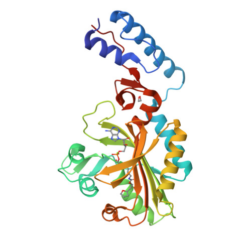
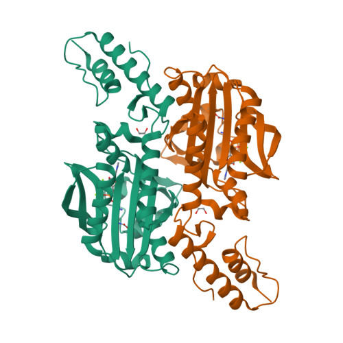
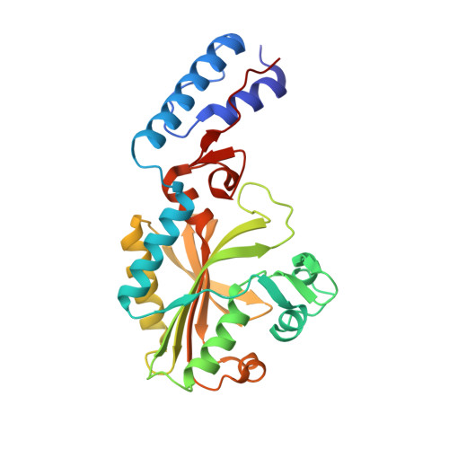
Structural Basis of Furan-Amino Acid Recognition by a Polyspecific Aminoacyl-tRNA-Synthetase and its Genetic Encoding in Human Cells.

Schmidt, M.J.Weber, A.Pott, M.Welte, W.Summerer, D.

(2014) Chembiochem 15: 1755

  • DOI: https://doi.org/10.1002/cbic.201402006
  • Primary Citation of Related Structures:  
    4CS2, 4CS3, 4CS4

  • PubMed Abstract: 

    The site-selective introduction of photo-crosslinking groups into proteins enables the discovery and mapping of weak and/or transient protein interactions with high spatiotemporal resolution, both in vitro and in vivo. We report the genetic encoding of a furan-based, photo-crosslinking amino acid in human cells; it can be activated with red light, thus offering high penetration depths in biological samples. This is achieved by activation of the amino acid and charging to its cognate tRNA by a pyrrolysyl-tRNA-synthetase (PylRS) mutant with broad polyspecificity. To gain insights into the recognition of this amino acid and to provide a rationale for its polyspecificity, we solved three crystal structures of the PylRS mutant: in its apo-form, in complex with adenosine 5'-(β,γ-imido)triphosphate (AMP-PNP) and in complex with the AMP ester of the furan amino acid. These structures provide clues for the observed polyspecificity and represent a promising starting point for the engineering of PylRS mutants with further increased substrate scope.


  • Organizational Affiliation

    Department of Chemistry, University of Konstanz, Universitätsstrasse 10, 78457 Konstanz (Germany).


Macromolecules
Find similar proteins by:  (by identity cutoff)  |  3D Structure
Entity ID: 1
MoleculeChains Sequence LengthOrganismDetailsImage
PYRROLYSINE--TRNA LIGASE274Methanosarcina mazeiMutation(s): 2 
EC: 6.1.1.26
UniProt
Find proteins for Q8PWY1 (Methanosarcina mazei (strain ATCC BAA-159 / DSM 3647 / Goe1 / Go1 / JCM 11833 / OCM 88))
Explore Q8PWY1 
Go to UniProtKB:  Q8PWY1
Entity Groups  
Sequence Clusters30% Identity50% Identity70% Identity90% Identity95% Identity100% Identity
UniProt GroupQ8PWY1
Sequence Annotations
Expand
  • Reference Sequence
Small Molecules
Ligands 6 Unique
IDChains Name / Formula / InChI Key2D Diagram3D Interactions
AMP
Query on AMP

Download Ideal Coordinates CCD File 
C [auth A]ADENOSINE MONOPHOSPHATE
C10 H14 N5 O7 P
UDMBCSSLTHHNCD-KQYNXXCUSA-N
POP
Query on POP

Download Ideal Coordinates CCD File 
E [auth A]PYROPHOSPHATE 2-
H2 O7 P2
XPPKVPWEQAFLFU-UHFFFAOYSA-L
FU0
Query on FU0

Download Ideal Coordinates CCD File 
D [auth A]2-(furan-2-yl)ethyl hydrogen carbonate
C7 H8 O4
SFUJACCQACPZEZ-UHFFFAOYSA-N
LYS
Query on LYS

Download Ideal Coordinates CCD File 
B [auth A]LYSINE
C6 H15 N2 O2
KDXKERNSBIXSRK-YFKPBYRVSA-O
EDO
Query on EDO

Download Ideal Coordinates CCD File 
H [auth A]1,2-ETHANEDIOL
C2 H6 O2
LYCAIKOWRPUZTN-UHFFFAOYSA-N
MG
Query on MG

Download Ideal Coordinates CCD File 
F [auth A],
G [auth A]
MAGNESIUM ION
Mg
JLVVSXFLKOJNIY-UHFFFAOYSA-N
Experimental Data & Validation

Experimental Data

  • Method: X-RAY DIFFRACTION
  • Resolution: 1.50 Å
  • R-Value Free:  0.185 (Depositor), 0.190 (DCC) 
  • R-Value Work:  0.155 (Depositor), 0.160 (DCC) 
  • R-Value Observed: 0.157 (Depositor) 
Space Group: C 1 2 1
Unit Cell:
Length ( Å )Angle ( ˚ )
a = 102.335α = 90
b = 44.345β = 99.85
c = 64.165γ = 90
Software Package:
Software NamePurpose
PHENIXrefinement
XDSdata reduction
PHENIXphasing

Structure Validation

View Full Validation Report


Ligand Structure Quality Assessment 

Created with Raphaël 2.3.0Worse 01 BetterLigand structure goodness of fit to experimental dataBest fitted AMPClick on this verticalbar to view detailsBest fitted FU0Click on this verticalbar to view details

Entry History 

Deposition Data

Revision History  (Full details and data files)

  • Version 1.0: 2014-04-30
    Type: Initial release
  • Version 1.1: 2014-08-27
    Changes: Database references
  • Version 1.2: 2024-05-08
    Changes: Data collection, Database references, Derived calculations, Other