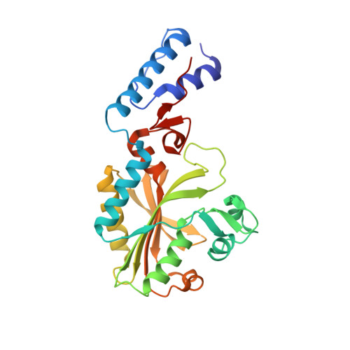
4CS4

Catalytic domain of Pyrrolysyl-tRNA synthetase mutant Y306A, Y384F in complex with AMPPNP


Experimental Data Snapshot

  • Method: X-RAY DIFFRACTION
  • Resolution: 1.35 Å
  • R-Value Free: 0.176 
  • R-Value Work: 0.148 
  • R-Value Observed: 0.150 

wwPDB Validation   3D Report Full Report


Ligand Structure Quality Assessment 


This is version 1.3 of the entry. See complete history


Literature

Structural Basis of Furan-Amino Acid Recognition by a Polyspecific Aminoacyl-tRNA-Synthetase and its Genetic Encoding in Human Cells.

Schmidt, M.J.Weber, A.Pott, M.Welte, W.Summerer, D.

(2014) Chembiochem 15: 1755

  • DOI: https://doi.org/10.1002/cbic.201402006
  • Primary Citation of Related Structures:  
    4CS2, 4CS3, 4CS4

  • PubMed Abstract: 

    The site-selective introduction of photo-crosslinking groups into proteins enables the discovery and mapping of weak and/or transient protein interactions with high spatiotemporal resolution, both in vitro and in vivo. We report the genetic encoding of a furan-based, photo-crosslinking amino acid in human cells; it can be activated with red light, thus offering high penetration depths in biological samples. This is achieved by activation of the amino acid and charging to its cognate tRNA by a pyrrolysyl-tRNA-synthetase (PylRS) mutant with broad polyspecificity. To gain insights into the recognition of this amino acid and to provide a rationale for its polyspecificity, we solved three crystal structures of the PylRS mutant: in its apo-form, in complex with adenosine 5'-(β,γ-imido)triphosphate (AMP-PNP) and in complex with the AMP ester of the furan amino acid. These structures provide clues for the observed polyspecificity and represent a promising starting point for the engineering of PylRS mutants with further increased substrate scope.


  • Organizational Affiliation

    Department of Chemistry, University of Konstanz, Universitätsstrasse 10, 78457 Konstanz (Germany).


Macromolecules
Find similar proteins by:  (by identity cutoff)  |  3D Structure
Entity ID: 1
MoleculeChains Sequence LengthOrganismDetailsImage
PYRROLYSYL-TRNA SYNTHETASE274Methanosarcina mazeiMutation(s): 2 
EC: 6.1.1.26
UniProt
Find proteins for Q8PWY1 (Methanosarcina mazei (strain ATCC BAA-159 / DSM 3647 / Goe1 / Go1 / JCM 11833 / OCM 88))
Explore Q8PWY1 
Go to UniProtKB:  Q8PWY1
Entity Groups  
Sequence Clusters30% Identity50% Identity70% Identity90% Identity95% Identity100% Identity
UniProt GroupQ8PWY1
Sequence Annotations
Expand
  • Reference Sequence
Small Molecules
Ligands 5 Unique
IDChains Name / Formula / InChI Key2D Diagram3D Interactions
ANP
Query on ANP

Download Ideal Coordinates CCD File 
B [auth A]PHOSPHOAMINOPHOSPHONIC ACID-ADENYLATE ESTER
C10 H17 N6 O12 P3
PVKSNHVPLWYQGJ-KQYNXXCUSA-N
AXZ
Query on AXZ

Download Ideal Coordinates CCD File 
Y [auth A]2-{[dihydroxy(4-aminoethylphenyl)-{4}-sulfanyl]amino}-3-hydroxypropanoic acid
C11 H18 N2 O5 S
SQBHFIANGBPIBX-JTQLQIEISA-N
PO4
Query on PO4

Download Ideal Coordinates CCD File 
Z [auth A]PHOSPHATE ION
O4 P
NBIIXXVUZAFLBC-UHFFFAOYSA-K
EDO
Query on EDO

Download Ideal Coordinates CCD File 
C [auth A]
D [auth A]
E [auth A]
F [auth A]
G [auth A]
C [auth A],
D [auth A],
E [auth A],
F [auth A],
G [auth A],
H [auth A],
I [auth A],
J [auth A],
K [auth A],
L [auth A],
M [auth A],
N [auth A],
O [auth A],
P [auth A],
Q [auth A],
R [auth A],
S [auth A],
T [auth A],
U [auth A],
V [auth A]
1,2-ETHANEDIOL
C2 H6 O2
LYCAIKOWRPUZTN-UHFFFAOYSA-N
MG
Query on MG

Download Ideal Coordinates CCD File 
W [auth A],
X [auth A]
MAGNESIUM ION
Mg
JLVVSXFLKOJNIY-UHFFFAOYSA-N
Experimental Data & Validation

Experimental Data

  • Method: X-RAY DIFFRACTION
  • Resolution: 1.35 Å
  • R-Value Free: 0.176 
  • R-Value Work: 0.148 
  • R-Value Observed: 0.150 
  • Space Group: C 1 2 1
Unit Cell:
Length ( Å )Angle ( ˚ )
a = 102.075α = 90
b = 44.533β = 99.66
c = 64.359γ = 90
Software Package:
Software NamePurpose
PHENIXrefinement
XDSdata reduction
PHENIXphasing

Structure Validation

View Full Validation Report


Ligand Structure Quality Assessment 


Entry History 

Deposition Data

Revision History  (Full details and data files)

  • Version 1.0: 2014-04-30
    Type: Initial release
  • Version 1.1: 2014-05-07
    Changes: Atomic model
  • Version 1.2: 2014-08-27
    Changes: Database references
  • Version 1.3: 2024-05-08
    Changes: Data collection, Database references, Derived calculations, Other