1MUQ | pdb_00001muq

X-ray Crystal Structure of Rattlesnake Venom Complexed With Thiodigalactoside


Experimental Data Snapshot

  • Method: X-RAY DIFFRACTION
  • Resolution: 2.30 Å
  • R-Value Free: 
    0.257 (Depositor), 0.260 (DCC) 
  • R-Value Work: 
    0.210 (Depositor), 0.210 (DCC) 
  • R-Value Observed: 
    0.210 (Depositor) 

wwPDB Validation   3D Report Full Report


Ligand Structure Quality Assessment 

Created with Raphaël 2.3.0Worse 01 BetterLigand structure goodness of fit to experimental dataBest fitted GALClick on this verticalbar to view details

This is version 3.1 of the entry. See complete history


Literature

X-ray Crystal Structure of a Galactose-Specific C-Type Lectin Possessing a Novel Decameric Quaternary Structure.

Walker, J.R.Nagar, B.Young, N.M.Hirama, T.Rini, J.M.

(2004) Biochemistry 43: 3783-3792

  • DOI: https://doi.org/10.1021/bi035871a
  • Primary Citation of Related Structures:  
    1JZN, 1MUQ

  • PubMed Abstract: 

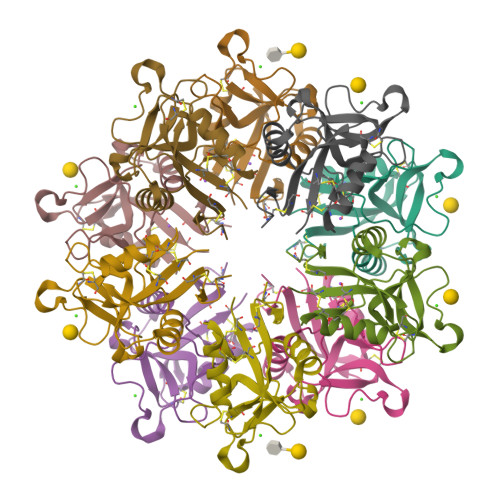
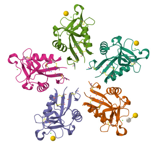
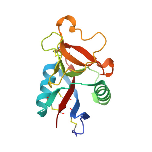
    Rattlesnake venom lectin (RSL) from the western diamondback rattlesnake (Crotalus atrox) is an oligomeric galactose-specific C-type lectin. The X-ray crystal structure of RSL, in complex with lactose and thiodigalactoside, at 2.2 and 2.3 A resolution, respectively, reveals a decameric protein composed of two 5-fold symmetric pentamers arranged in a staggered, back-to-back orientation. Each monomer corresponds to a single canonical C-type lectin carbohydrate recognition domain devoid of accessory domains and is disulfide-bonded to a monomer in the other pentamer. The structure is the first example of that of a carbohydrate complex of a vertebrate galactose-specific C-type lectin. The 10 carbohydrate-binding sites, located on the rim of the decamer, suggest a role for multivalent interactions and a mechanism for RSL's ability to promote receptor cross-linking and cell aggregation.


  • Organizational Affiliation

    Department of Molecular and Medical Genetics, University of Toronto, Toronto, Ontario M5S 1A8, Canada.


Macromolecules
Find similar proteins by:  (by identity cutoff)  |  3D Structure
Entity ID: 1
MoleculeChains Sequence LengthOrganismDetailsImage
Galactose-specific lectin
A, B, C, D, E
135Crotalus atroxMutation(s): 0 
UniProt
Find proteins for P21963 (Crotalus atrox)
Explore P21963 
Go to UniProtKB:  P21963
Entity Groups  
Sequence Clusters30% Identity50% Identity70% Identity90% Identity95% Identity100% Identity
UniProt GroupP21963
Sequence Annotations
Expand
  • Reference Sequence
Oligosaccharides

Help

Entity ID: 2
MoleculeChains Length2D Diagram Glycosylation3D Interactions
1-thio-beta-D-galactopyranose-(1-1)-beta-D-galactopyranoseF [auth G]2N/AN/A
Glycosylation Resources
GlyTouCan:  G61532BD
GlyCosmos:  G61532BD
Small Molecules
Ligands 3 Unique
IDChains Name / Formula / InChI Key2D Diagram3D Interactions
GAL
Query on GAL

Download Ideal Coordinates CCD File 
I [auth A],
N [auth C],
S [auth E]
beta-D-galactopyranose
C6 H12 O6
WQZGKKKJIJFFOK-FPRJBGLDSA-N
CA
Query on CA

Download Ideal Coordinates CCD File 
G [auth A],
J [auth B],
L [auth C],
O [auth D],
Q [auth E]
CALCIUM ION
Ca
BHPQYMZQTOCNFJ-UHFFFAOYSA-N
NA
Query on NA

Download Ideal Coordinates CCD File 
H [auth A],
K [auth B],
M [auth C],
P [auth D],
R [auth E]
SODIUM ION
Na
FKNQFGJONOIPTF-UHFFFAOYSA-N
Biologically Interesting Molecules (External Reference) 1 Unique
Experimental Data & Validation

Experimental Data

  • Method: X-RAY DIFFRACTION
  • Resolution: 2.30 Å
  • R-Value Free:  0.257 (Depositor), 0.260 (DCC) 
  • R-Value Work:  0.210 (Depositor), 0.210 (DCC) 
  • R-Value Observed: 0.210 (Depositor) 
Space Group: C 2 2 21
Unit Cell:
Length ( Å )Angle ( ˚ )
a = 110.454α = 90
b = 151.234β = 90
c = 95.014γ = 90
Software Package:
Software NamePurpose
DENZOdata reduction
SCALEPACKdata scaling
PHASESphasing
CNSrefinement

Structure Validation

View Full Validation Report


Ligand Structure Quality Assessment 

Created with Raphaël 2.3.0Worse 01 BetterLigand structure goodness of fit to experimental dataBest fitted GALClick on this verticalbar to view details

Entry History 

Deposition Data

Revision History  (Full details and data files)

  • Version 1.0: 2003-07-01
    Type: Initial release
  • Version 1.1: 2008-04-28
    Changes: Version format compliance
  • Version 1.2: 2011-07-13
    Changes: Derived calculations, Version format compliance
  • Version 2.0: 2020-07-29
    Type: Remediation
    Reason: Carbohydrate remediation
    Changes: Advisory, Atomic model, Data collection, Derived calculations, Non-polymer description, Structure summary
  • Version 3.0: 2020-10-14
    Changes: Atomic model, Data collection, Derived calculations, Structure summary
  • Version 3.1: 2024-11-20
    Changes: Data collection, Database references, Structure summary