3FBZ | pdb_00003fbz

Crystal structure of ORF140 of the archaeal virus Acidianus Filamentous Virus 1 (AFV1)


Experimental Data Snapshot

  • Method: X-RAY DIFFRACTION
  • Resolution: 2.30 Å
  • R-Value Free: 
    0.255 (Depositor), 0.270 (DCC) 
  • R-Value Work: 
    0.185 (Depositor), 0.200 (DCC) 
  • R-Value Observed: 
    0.188 (Depositor) 

wwPDB Validation   3D Report Full Report


Ligand Structure Quality Assessment 

Created with Raphaël 2.3.0Worse 01 BetterLigand structure goodness of fit to experimental dataBest fitted BOGClick on this verticalbar to view details

This is version 1.4 of the entry. See complete history


Literature

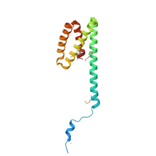
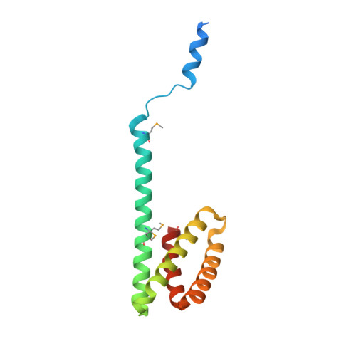
Acidianus filamentous virus 1 coat proteins display a helical fold spanning the filamentous archaeal viruses lineage.

Goulet, A.Blangy, S.Redder, P.Prangishvili, D.Felisberto-Rodrigues, C.Forterre, P.Campanacci, V.Cambillau, C.

(2009) Proc Natl Acad Sci U S A 106: 21155-21160

  • DOI: https://doi.org/10.1073/pnas.0909893106
  • Primary Citation of Related Structures:  
    3FBL, 3FBZ

  • PubMed Abstract: 

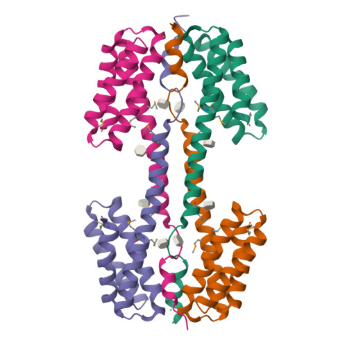
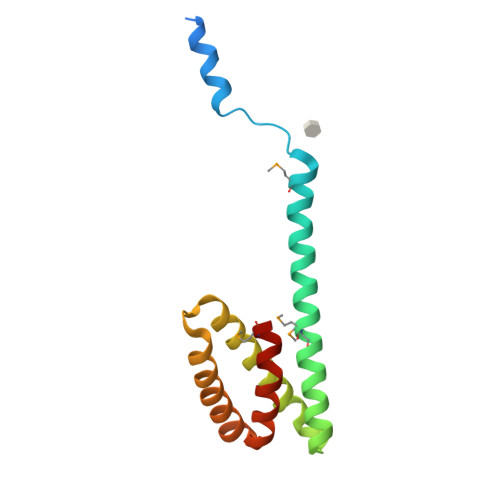
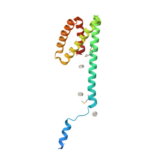
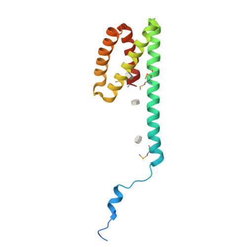
    Acidianus filamentous virus 1 (AFV1), a member of the Lipothrixviridae family, infects the hyperthermophilic, acidophilic crenarchaeaon Acidianus hospitalis. The virion, covered with a lipidic outer shell, is 9,100-A long and contains a 20.8-kb linear dsDNA genome. We have identified the two major coat proteins of the virion (MCPs; 132 and 140 amino acids). They bind DNA and form filaments when incubated with linear dsDNA. A C-terminal domain is identified in their crystal structure with a four-helix-bundle fold. In the topological model of the virion filament core, the genomic dsDNA superhelix wraps around the AFV1-132 basic protein, and the AFV1-140 basic N terminus binds genomic DNA, while its lipophilic C-terminal domain is imbedded in the lipidic outer shell. The four-helix bundle fold of the MCPs from AFV1 is identical to that of the coat protein (CP) of Sulfolobus islandicus rod-shaped virus (SIRV), a member of the Rudiviridae family. Despite low sequence identity between these proteins, their high degree of structural similarity suggests that they could have derived from a common ancestor and could thus define an yet undescribed viral lineage.


  • Organizational Affiliation

    Architecture et Fonction des Macromolécules Biologiques, Centre national de la Recherche Scientifique and Universités Aix-Marseille I & II, Architecture et Fonction des Macromolécules Biologiques, Unité Mixte de Recherche 6098, Case 932, 163 avenue de Luminy, 13288 Marseille Cedex 9, France.


Macromolecules
Find similar proteins by:  (by identity cutoff)  |  3D Structure
Entity ID: 1
MoleculeChains Sequence LengthOrganismDetailsImage
Putative uncharacterized protein
A, B, C, D
140Captovirus AFV1Mutation(s): 0 
Gene Names: AFV1_ORF140
UniProt
Find proteins for Q70LC6 (Acidianus filamentous virus 1 (isolate United States/Yellowstone))
Explore Q70LC6 
Go to UniProtKB:  Q70LC6
Entity Groups  
Sequence Clusters30% Identity50% Identity70% Identity90% Identity95% Identity100% Identity
UniProt GroupQ70LC6
Sequence Annotations
Expand
  • Reference Sequence
Small Molecules
Ligands 2 Unique
IDChains Name / Formula / InChI Key2D Diagram3D Interactions
BOG
Query on BOG

Download Ideal Coordinates CCD File 
E [auth A]
F [auth B]
G [auth B]
H [auth B]
J [auth C]
octyl beta-D-glucopyranoside
C14 H28 O6
HEGSGKPQLMEBJL-RKQHYHRCSA-N
CL
Query on CL

Download Ideal Coordinates CCD File 
I [auth B],
N [auth D]
CHLORIDE ION
Cl
VEXZGXHMUGYJMC-UHFFFAOYSA-M
Modified Residues  1 Unique
IDChains TypeFormula2D DiagramParent
MSE
Query on MSE
A, B, C, D
L-PEPTIDE LINKINGC5 H11 N O2 SeMET
Experimental Data & Validation

Experimental Data

  • Method: X-RAY DIFFRACTION
  • Resolution: 2.30 Å
  • R-Value Free:  0.255 (Depositor), 0.270 (DCC) 
  • R-Value Work:  0.185 (Depositor), 0.200 (DCC) 
  • R-Value Observed: 0.188 (Depositor) 
Space Group: P 21 21 21
Unit Cell:
Length ( Å )Angle ( ˚ )
a = 58.158α = 90
b = 81.15β = 90
c = 119.898γ = 90
Software Package:
Software NamePurpose
SHELXSphasing
REFMACrefinement
MOSFLMdata reduction
SCALAdata scaling

Structure Validation

View Full Validation Report


Ligand Structure Quality Assessment 

Created with Raphaël 2.3.0Worse 01 BetterLigand structure goodness of fit to experimental dataBest fitted BOGClick on this verticalbar to view details

Entry History 

Deposition Data

Revision History  (Full details and data files)

  • Version 1.0: 2009-11-10
    Type: Initial release
  • Version 1.1: 2011-07-13
    Changes: Version format compliance
  • Version 1.2: 2020-07-29
    Type: Remediation
    Reason: Carbohydrate remediation
    Changes: Data collection, Database references, Derived calculations, Structure summary
  • Version 1.3: 2023-12-27
    Changes: Data collection, Database references, Structure summary
  • Version 1.4: 2024-11-20
    Changes: Structure summary