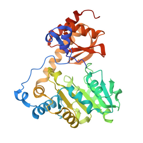
9JFB

Crystal structure of L-threonine-O-3-phosphate decarboxylase CobC


Experimental Data Snapshot

  • Method: X-RAY DIFFRACTION
  • Resolution: 2.20 Å
  • R-Value Free: 0.185 
  • R-Value Work: 0.154 
  • R-Value Observed: 0.156 

Starting Model: in silico
View more details

wwPDB Validation   3D Report Full Report


This is version 1.0 of the entry. See complete history


Literature

Crystal structure of l-threonine-O-3-phosphate decarboxylase CobC from Sinorhizobium meliloti involved in vitamin B 12 biosynthesis.

Jiang, M.Guo, S.Chen, X.Wei, Q.Wang, M.

(2024) Biochem Biophys Res Commun 734: 150767-150767

  • DOI: https://doi.org/10.1016/j.bbrc.2024.150767
  • Primary Citation of Related Structures:  
    9JFB, 9JFF

  • PubMed Abstract: 

    Vitamin B 12 is involved in many important biochemical reactions for humans, and its deficiency can lead to serious diseases. The industrial production of vitamin B 12 is achieved through microbial fermentation. In this work, we determine the crystal structures of the l-threonine-O-3-phosphate (Thr-P) decarboxylase CobC from Sinorhizobium meliloti (SmCobC), an industrial vitamin B 12 -producing bacterium, in apo form and in complex with a reaction intermediate. Our structures supported the Thr-P decarboxylase activity of SmCobC and revealed that the positively charged substrate-binding pocket between the large and small domains determines its substrate selectivity for Thr-P. Moreover, our results provided evidence for the proposition that the AP-P linker is formed by direct incorporation of AP-P in the biosynthetic pathway of vitamin B 12 in S.meliloti.


  • Organizational Affiliation

    Institutes of Physical Science and Information Technology, Anhui University, Hefei, 230601, Anhui, China; School of Life Sciences, Anhui University, Hefei, 230601, Anhui, China.


Macromolecules
Find similar proteins by:  (by identity cutoff)  |  3D Structure
Entity ID: 1
MoleculeChains Sequence LengthOrganismDetailsImage
threonine-phosphate decarboxylase341Sinorhizobium melilotiMutation(s): 0 
Gene Names: cobC
EC: 4.1.1.81
UniProt
Find proteins for A0A499W357 (Rhizobium meliloti)
Explore A0A499W357 
Go to UniProtKB:  A0A499W357
Entity Groups  
Sequence Clusters30% Identity50% Identity70% Identity90% Identity95% Identity100% Identity
UniProt GroupA0A499W357
Sequence Annotations
Expand
  • Reference Sequence
Small Molecules
Ligands 4 Unique
IDChains Name / Formula / InChI Key2D Diagram3D Interactions
SO4
Query on SO4

Download Ideal Coordinates CCD File 
E [auth A]
F [auth A]
G [auth A]
H [auth A]
I [auth A]
E [auth A],
F [auth A],
G [auth A],
H [auth A],
I [auth A],
J [auth A],
K [auth A],
L [auth A],
M [auth A]
SULFATE ION
O4 S
QAOWNCQODCNURD-UHFFFAOYSA-L
GOL
Query on GOL

Download Ideal Coordinates CCD File 
B [auth A],
C [auth A],
D [auth A]
GLYCEROL
C3 H8 O3
PEDCQBHIVMGVHV-UHFFFAOYSA-N
CL
Query on CL

Download Ideal Coordinates CCD File 
N [auth A],
O [auth A],
P [auth A],
Q [auth A]
CHLORIDE ION
Cl
VEXZGXHMUGYJMC-UHFFFAOYSA-M
NA
Query on NA

Download Ideal Coordinates CCD File 
R [auth A]SODIUM ION
Na
FKNQFGJONOIPTF-UHFFFAOYSA-N
Experimental Data & Validation

Experimental Data

  • Method: X-RAY DIFFRACTION
  • Resolution: 2.20 Å
  • R-Value Free: 0.185 
  • R-Value Work: 0.154 
  • R-Value Observed: 0.156 
  • Space Group: I 21 3
Unit Cell:
Length ( Å )Angle ( ˚ )
a = 159.418α = 90
b = 159.418β = 90
c = 159.418γ = 90
Software Package:
Software NamePurpose
PHENIXrefinement
HKL-3000data scaling
HKL-3000data reduction
PHASERphasing

Structure Validation

View Full Validation Report


Entry History & Funding Information

Deposition Data


Funding OrganizationLocationGrant Number
Other governmentChina2108085MC75

Revision History  (Full details and data files)

  • Version 1.0: 2024-10-16
    Type: Initial release