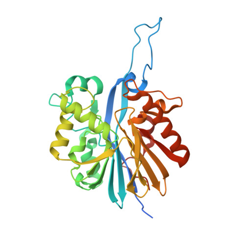
9AYT

Structure of the quorum quenching lactonase GcL bound to N-hexanoyl-L-homoserine lactone


Experimental Data Snapshot

  • Method: X-RAY DIFFRACTION
  • Resolution: 2.10 Å
  • R-Value Free: 0.220 
  • R-Value Work: 0.192 
  • R-Value Observed: 0.194 

Starting Model: experimental
View more details

wwPDB Validation   3D Report Full Report


Ligand Structure Quality Assessment 


This is version 1.0 of the entry. See complete history


Literature

Catalytic Redundancies and Conformational Plasticity Drives Selectivity and Promiscuity in Quorum Quenching Lactonases.

Corbella, M.Bravo, J.Demkiv, A.O.Calixto, A.R.Sompiyachoke, K.Bergonzi, C.Brownless, A.R.Elias, M.H.Kamerlin, S.C.L.

(2024) JACS Au 4: 3519-3536

  • DOI: https://doi.org/10.1021/jacsau.4c00404
  • Primary Citation of Related Structures:  
    9AYT, 9B2I, 9B2J, 9B2L, 9B2N, 9B2O, 9B2P

  • PubMed Abstract: 

    Several enzymes from the metallo-β-lactamase-like family of lactonases (MLLs) degrade N- acyl L-homoserine lactones (AHLs). They play a role in a microbial communication system known as quorum sensing, which contributes to pathogenicity and biofilm formation. Designing quorum quenching ( QQ ) enzymes that can interfere with this communication allows them to be used in a range of industrial and biomedical applications. However, tailoring these enzymes for specific communication signals requires a thorough understanding of their mechanisms and the physicochemical properties that determine their substrate specificities. We present here a detailed biochemical, computational, and structural study of GcL, which is a highly proficient and thermostable MLL with broad substrate specificity. We show that GcL not only accepts a broad range of substrates but also hydrolyzes these substrates through at least two different mechanisms. Further, the preferred mechanism appears to depend on both the substrate structure and/or the nature of the residues lining the active site. We demonstrate that other lactonases, such as AiiA and AaL, show similar mechanistic promiscuity, suggesting that this is a shared feature among MLLs. Mechanistic promiscuity has been seen previously in the lactonase/paraoxonase PON1, as well as with protein tyrosine phosphatases that operate via a dual general acid mechanism. The apparent prevalence of this phenomenon is significant from both a biochemical and protein engineering perspective: in addition to optimizing for specific substrates, it may be possible to optimize for specific mechanisms, opening new doors not just for the design of novel quorum quenching enzymes but also of other mechanistically promiscuous enzymes.


  • Organizational Affiliation

    Departament de Química Inorgànica (Seeió de Química Orgànica) & Institut de Química Teòrica i Computacional (IQTCUB), Universitat de Barcelona, Martíi Franquès 1, 08028 Barcelona, Spain.


Macromolecules
Find similar proteins by:  (by identity cutoff)  |  3D Structure
Entity ID: 1
MoleculeChains Sequence LengthOrganismDetailsImage
GcL lactonaseA [auth P],
B [auth D]
297Parageobacillus caldoxylosilyticusMutation(s): 0 
Gene Names: GCA01S_030_00190
UniProt
Find proteins for A0A023DFE8 (Parageobacillus caldoxylosilyticus NBRC 107762)
Explore A0A023DFE8 
Go to UniProtKB:  A0A023DFE8
Entity Groups  
Sequence Clusters30% Identity50% Identity70% Identity90% Identity95% Identity100% Identity
UniProt GroupA0A023DFE8
Sequence Annotations
Expand
  • Reference Sequence
Small Molecules
Ligands 7 Unique
IDChains Name / Formula / InChI Key2D Diagram3D Interactions
1PE
Query on 1PE

Download Ideal Coordinates CCD File 
R [auth P]PENTAETHYLENE GLYCOL
C10 H22 O6
JLFNLZLINWHATN-UHFFFAOYSA-N
HL6 (Subject of Investigation/LOI)
Query on HL6

Download Ideal Coordinates CCD File 
KA [auth D],
Q [auth P]
N-[(3S)-2-oxotetrahydrofuran-3-yl]hexanamide
C10 H17 N O3
ZJFKKPDLNLCPNP-QMMMGPOBSA-N
SO4
Query on SO4

Download Ideal Coordinates CCD File 
LA [auth D],
S [auth P]
SULFATE ION
O4 S
QAOWNCQODCNURD-UHFFFAOYSA-L
EDO
Query on EDO

Download Ideal Coordinates CCD File 
AA [auth D]
BA [auth D]
CA [auth D]
DA [auth D]
E [auth P]
AA [auth D],
BA [auth D],
CA [auth D],
DA [auth D],
E [auth P],
F [auth P],
G [auth P],
H [auth P],
I [auth P],
J [auth P],
K [auth P],
L [auth P],
M [auth P],
V [auth D],
W [auth D],
X [auth D],
Y [auth D],
Z [auth D]
1,2-ETHANEDIOL
C2 H6 O2
LYCAIKOWRPUZTN-UHFFFAOYSA-N
ACT
Query on ACT

Download Ideal Coordinates CCD File 
EA [auth D]
FA [auth D]
GA [auth D]
HA [auth D]
IA [auth D]
EA [auth D],
FA [auth D],
GA [auth D],
HA [auth D],
IA [auth D],
JA [auth D],
N [auth P],
O [auth P],
P
ACETATE ION
C2 H3 O2
QTBSBXVTEAMEQO-UHFFFAOYSA-M
CO
Query on CO

Download Ideal Coordinates CCD File 
C [auth P],
T [auth D]
COBALT (II) ION
Co
XLJKHNWPARRRJB-UHFFFAOYSA-N
FE
Query on FE

Download Ideal Coordinates CCD File 
D [auth P],
U [auth D]
FE (III) ION
Fe
VTLYFUHAOXGGBS-UHFFFAOYSA-N
Experimental Data & Validation

Experimental Data

  • Method: X-RAY DIFFRACTION
  • Resolution: 2.10 Å
  • R-Value Free: 0.220 
  • R-Value Work: 0.192 
  • R-Value Observed: 0.194 
  • Space Group: H 3
Unit Cell:
Length ( Å )Angle ( ˚ )
a = 108.5α = 90
b = 108.5β = 90
c = 228.5γ = 120
Software Package:
Software NamePurpose
REFMACrefinement
XDSdata reduction
XSCALEdata scaling
REFMACphasing

Structure Validation

View Full Validation Report


Ligand Structure Quality Assessment 


Entry History & Funding Information

Deposition Data


Funding OrganizationLocationGrant Number
National Institutes of Health/National Institute of General Medical Sciences (NIH/NIGMS)United StatesR35GM133487

Revision History  (Full details and data files)

  • Version 1.0: 2024-11-06
    Type: Initial release