8Q66 | pdb_00008q66

Crystal Structure of the C. elegans MUT-7 MUT-8 CTD complex


Experimental Data Snapshot

  • Method: X-RAY DIFFRACTION
  • Resolution: 2.03 Å
  • R-Value Free: 
    0.225 (Depositor), 0.220 (DCC) 
  • R-Value Work: 
    0.191 (Depositor), 0.190 (DCC) 
  • R-Value Observed: 
    0.193 (Depositor) 

Starting Model: in silico
View more details

wwPDB Validation   3D Report Full Report


This is version 1.1 of the entry. See complete history


Literature

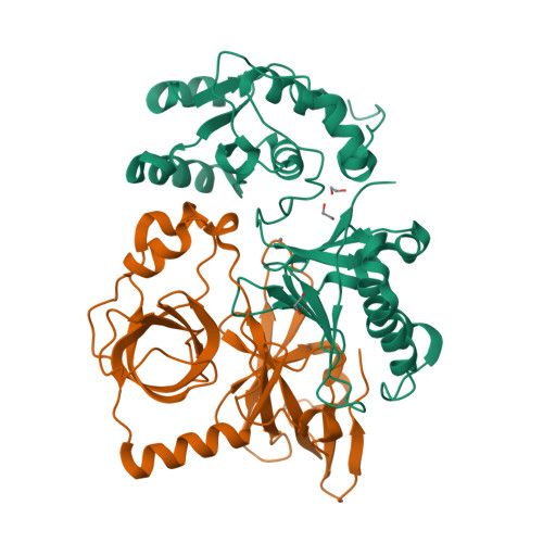
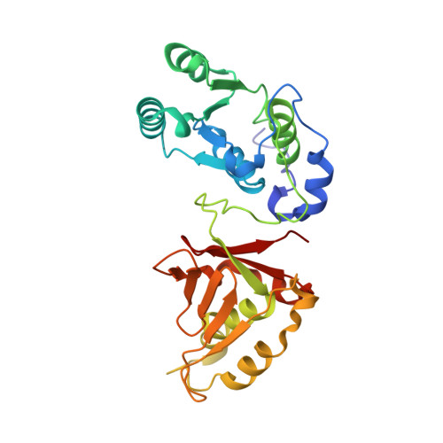
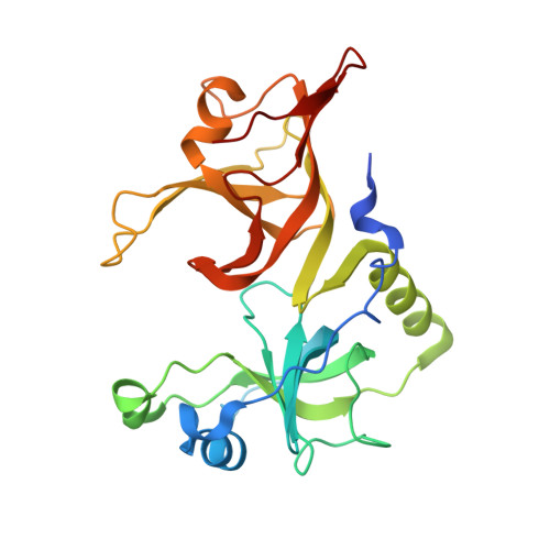
MUT-7 exoribonuclease activity and localization are mediated by an ancient domain.

Busetto, V.Pshanichnaya, L.Lichtenberger, R.Hann, S.Ketting, R.F.Falk, S.

(2024) Nucleic Acids Res 52: 9076-9091

  • DOI: https://doi.org/10.1093/nar/gkae610
  • Primary Citation of Related Structures:  
    8Q66

  • PubMed Abstract: 

    The MUT-7 family of 3'-5' exoribonucleases is evolutionarily conserved across the animal kingdom and plays essential roles in small RNA production in the germline. Most MUT-7 homologues carry a C-terminal domain of unknown function named MUT7-C appended to the exoribonuclease domain. Our analysis shows that the MUT7-C is evolutionary ancient, as a minimal version of the domain exists as an individual protein in prokaryotes. In animals, MUT7-C has acquired an insertion that diverged during evolution, expanding its functions. Caenorhabditis elegans MUT-7 contains a specific insertion within MUT7-C, which allows binding to MUT-8 and, consequently, MUT-7 recruitment to germ granules. In addition, in C. elegans and human MUT-7, the MUT7-C domain contributes to RNA binding and is thereby crucial for ribonuclease activity. This RNA-binding function most likely represents the ancestral function of the MUT7-C domain. Overall, this study sheds light on MUT7-C and assigns two functions to this previously uncharacterized domain.


  • Organizational Affiliation

    Max Perutz Labs, Vienna Biocenter Campus (VBC), Dr.-Bohr-Gasse 9, 1030 Vienna, Austria.


Macromolecules
Find similar proteins by:  (by identity cutoff)  |  3D Structure
Entity ID: 1
MoleculeChains Sequence LengthOrganismDetailsImage
Exonuclease mut-7272Caenorhabditis elegansMutation(s): 0 
Gene Names: mut-7
EC: 3.1
UniProt
Find proteins for P34607 (Caenorhabditis elegans)
Explore P34607 
Go to UniProtKB:  P34607
Entity Groups  
Sequence Clusters30% Identity50% Identity70% Identity90% Identity95% Identity100% Identity
UniProt GroupP34607
Sequence Annotations
Expand
  • Reference Sequence
Find similar proteins by:  (by identity cutoff)  |  3D Structure
Entity ID: 2
MoleculeChains Sequence LengthOrganismDetailsImage
SH2 domain-containing protein251Caenorhabditis elegansMutation(s): 0 
Gene Names: rde-2
UniProt
Find proteins for Q19672 (Caenorhabditis elegans)
Explore Q19672 
Go to UniProtKB:  Q19672
Entity Groups  
Sequence Clusters30% Identity50% Identity70% Identity90% Identity95% Identity100% Identity
UniProt GroupQ19672
Sequence Annotations
Expand
  • Reference Sequence
Experimental Data & Validation

Experimental Data

  • Method: X-RAY DIFFRACTION
  • Resolution: 2.03 Å
  • R-Value Free:  0.225 (Depositor), 0.220 (DCC) 
  • R-Value Work:  0.191 (Depositor), 0.190 (DCC) 
  • R-Value Observed: 0.193 (Depositor) 
Space Group: C 1 2 1
Unit Cell:
Length ( Å )Angle ( ˚ )
a = 145.089α = 90
b = 59.053β = 119.8
c = 88.161γ = 90
Software Package:
Software NamePurpose
PHENIXphasing
PHENIXrefinement
PDB-REDOrefinement
pointlessdata scaling
XDSdata reduction

Structure Validation

View Full Validation Report


Entry History & Funding Information

Deposition Data


Funding OrganizationLocationGrant Number
H2020 Marie Curie Actions of the European CommissionEuropean UnionFA743031

Revision History  (Full details and data files)

  • Version 1.0: 2024-03-06
    Type: Initial release
  • Version 1.1: 2024-09-18
    Changes: Database references