8GZW | pdb_00008gzw

Klebsiella pneumoniae FtsZ complexed with monobody (P21)


Experimental Data Snapshot

  • Method: X-RAY DIFFRACTION
  • Resolution: 2.50 Å
  • R-Value Free: 
    0.284 (Depositor), 0.280 (DCC) 
  • R-Value Work: 
    0.215 (Depositor), 0.210 (DCC) 
  • R-Value Observed: 
    0.218 (Depositor) 

Starting Model: experimental
View more details

wwPDB Validation   3D Report Full Report


Ligand Structure Quality Assessment 

Created with Raphaël 2.3.0Worse 01 BetterLigand structure goodness of fit to experimental dataBest fitted GDPClick on this verticalbar to view details

This is version 1.1 of the entry. See complete history


Literature

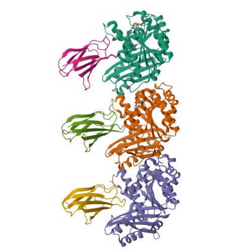
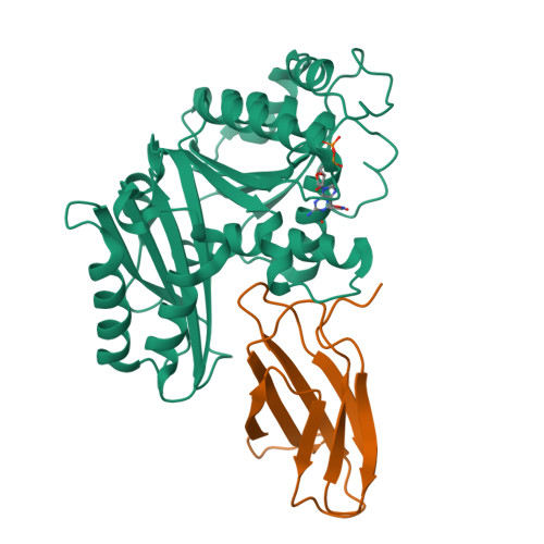
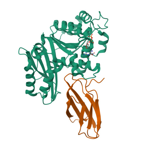
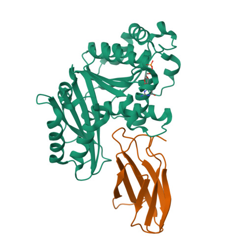
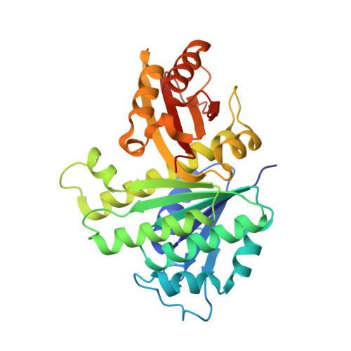
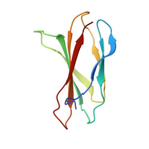
Structures of a FtsZ single protofilament and a double-helical tube in complex with a monobody.

Fujita, J.Amesaka, H.Yoshizawa, T.Hibino, K.Kamimura, N.Kuroda, N.Konishi, T.Kato, Y.Hara, M.Inoue, T.Namba, K.Tanaka, S.I.Matsumura, H.

(2023) Nat Commun 14: 4073-4073

  • DOI: https://doi.org/10.1038/s41467-023-39807-5
  • Primary Citation of Related Structures:  
    8GZV, 8GZW, 8GZX, 8GZY, 8H1O, 8IBN

  • PubMed Abstract: 

    FtsZ polymerizes into protofilaments to form the Z-ring that acts as a scaffold for accessory proteins during cell division. Structures of FtsZ have been previously solved, but detailed mechanistic insights are lacking. Here, we determine the cryoEM structure of a single protofilament of FtsZ from Klebsiella pneumoniae (KpFtsZ) in a polymerization-preferred conformation. We also develop a monobody (Mb) that binds to KpFtsZ and FtsZ from Escherichia coli without affecting their GTPase activity. Crystal structures of the FtsZ-Mb complexes reveal the Mb binding mode, while addition of Mb in vivo inhibits cell division. A cryoEM structure of a double-helical tube of KpFtsZ-Mb at 2.7 Å resolution shows two parallel protofilaments. Our present study highlights the physiological roles of the conformational changes of FtsZ in treadmilling that regulate cell division.


  • Organizational Affiliation

    Graduate School of Frontier Biosciences, Osaka University, 1-3 Yamadaoka, Suita, Osaka, 565-0871, Japan.


Macromolecules
Find similar proteins by:  (by identity cutoff)  |  3D Structure
Entity ID: 1
MoleculeChains Sequence LengthOrganismDetailsImage
Cell division protein FtsZ
A, C, E
309Klebsiella pneumoniaeMutation(s): 0 
Gene Names: ftsZ
UniProt
Find proteins for A0A0H3GRR0 (Klebsiella pneumoniae subsp. pneumoniae (strain HS11286))
Explore A0A0H3GRR0 
Go to UniProtKB:  A0A0H3GRR0
Entity Groups  
Sequence Clusters30% Identity50% Identity70% Identity90% Identity95% Identity100% Identity
UniProt GroupA0A0H3GRR0
Sequence Annotations
Expand
  • Reference Sequence
Find similar proteins by:  (by identity cutoff)  |  3D Structure
Entity ID: 2
MoleculeChains Sequence LengthOrganismDetailsImage
Monobody
B, D, F
92Homo sapiensMutation(s): 0 
Entity Groups  
Sequence Clusters30% Identity50% Identity70% Identity90% Identity95% Identity100% Identity
Sequence Annotations
Expand
  • Reference Sequence
Experimental Data & Validation

Experimental Data

  • Method: X-RAY DIFFRACTION
  • Resolution: 2.50 Å
  • R-Value Free:  0.284 (Depositor), 0.280 (DCC) 
  • R-Value Work:  0.215 (Depositor), 0.210 (DCC) 
  • R-Value Observed: 0.218 (Depositor) 
Space Group: P 1 21 1
Unit Cell:
Length ( Å )Angle ( ˚ )
a = 88.471α = 90
b = 66.933β = 92.055
c = 102.172γ = 90
Software Package:
Software NamePurpose
PHENIXrefinement
XDSdata reduction
XDSdata scaling
MOLREPphasing

Structure Validation

View Full Validation Report


Ligand Structure Quality Assessment 

Created with Raphaël 2.3.0Worse 01 BetterLigand structure goodness of fit to experimental dataBest fitted GDPClick on this verticalbar to view details

Entry History & Funding Information

Deposition Data


Funding OrganizationLocationGrant Number
Japan Society for the Promotion of Science (JSPS)JapanJP18K06094

Revision History  (Full details and data files)

  • Version 1.0: 2023-07-19
    Type: Initial release
  • Version 1.1: 2023-11-29
    Changes: Data collection, Refinement description