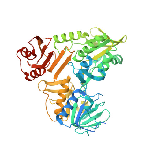
8D3O

Crystal structure of human Apoptosis-Inducing Factor (AIF) complexed with 8-methoxyquinolin-4-amine


Experimental Data Snapshot

  • Method: X-RAY DIFFRACTION
  • Resolution: 2.25 Å
  • R-Value Free: 0.237 
  • R-Value Work: 0.213 
  • R-Value Observed: 0.214 

wwPDB Validation   3D Report Full Report


Ligand Structure Quality Assessment 


This is version 1.1 of the entry. See complete history


Literature

Chemical screening by time-resolved X-ray scattering to discover allosteric probes.

Brosey, C.A.Link, T.M.Shen, R.Moiani, D.Burnett, K.Hura, G.L.Jones, D.E.Tainer, J.A.

(2024) Nat Chem Biol 

  • DOI: https://doi.org/10.1038/s41589-024-01609-1
  • Primary Citation of Related Structures:  
    8D3E, 8D3G, 8D3H, 8D3I, 8D3J, 8D3K, 8D3N, 8D3O

  • PubMed Abstract: 

    Drug discovery relies on efficient identification of small-molecule leads and their interactions with macromolecular targets. However, understanding how chemotypes impact mechanistically important conformational states often remains secondary among high-throughput discovery methods. Here, we present a conformational discovery pipeline integrating time-resolved, high-throughput small-angle X-ray scattering (TR-HT-SAXS) and classic fragment screening applied to allosteric states of the mitochondrial import oxidoreductase apoptosis-inducing factor (AIF). By monitoring oxidized and X-ray-reduced AIF states, TR-HT-SAXS leverages structure and kinetics to generate a multidimensional screening dataset that identifies fragment chemotypes allosterically stimulating AIF dimerization. Fragment-induced dimerization rates, quantified with time-resolved SAXS similarity analysis (k VR ), capture structure-activity relationships (SAR) across the top-ranked 4-aminoquinoline chemotype. Crystallized AIF-aminoquinoline complexes validate TR-SAXS-guided SAR, supporting this conformational chemotype for optimization. AIF-aminoquinoline structures and mutational analysis reveal active site F482 as an underappreciated allosteric stabilizer of AIF dimerization. This conformational discovery pipeline illustrates TR-HT-SAXS as an effective technology for targeting chemical leads to important macromolecular states.


  • Organizational Affiliation

    Department of Molecular and Cellular Oncology, The University of Texas MD Anderson Cancer Center, Houston, TX, USA. [email protected].


Macromolecules
Find similar proteins by:  (by identity cutoff)  |  3D Structure
Entity ID: 1
MoleculeChains Sequence LengthOrganismDetailsImage
Apoptosis-inducing factor 1, mitochondrial
A, B
543Homo sapiensMutation(s): 0 
Gene Names: AIFM1AIFPDCD8
EC: 1.6.99
UniProt & NIH Common Fund Data Resources
Find proteins for O95831 (Homo sapiens)
Explore O95831 
Go to UniProtKB:  O95831
PHAROS:  O95831
GTEx:  ENSG00000156709 
Entity Groups  
Sequence Clusters30% Identity50% Identity70% Identity90% Identity95% Identity100% Identity
UniProt GroupO95831
Sequence Annotations
Expand
  • Reference Sequence

Find similar proteins by:  Sequence   |   3D Structure  

Entity ID: 2
MoleculeChains Sequence LengthOrganismDetailsImage
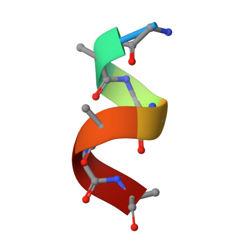
Apoptosis-inducing factor (AIF), unidentified helixC [auth D]7Homo sapiensMutation(s): 0 
Gene Names: AIFM1
EC: 1.6.99
Sequence Annotations
Expand
  • Reference Sequence
Small Molecules
Ligands 4 Unique
IDChains Name / Formula / InChI Key2D Diagram3D Interactions
FAD
Query on FAD

Download Ideal Coordinates CCD File 
F [auth A],
K [auth B]
FLAVIN-ADENINE DINUCLEOTIDE
C27 H33 N9 O15 P2
VWWQXMAJTJZDQX-UYBVJOGSSA-N
QF6 (Subject of Investigation/LOI)
Query on QF6

Download Ideal Coordinates CCD File 
D [auth A],
J [auth B]
8-methoxyquinolin-4-amine
C10 H10 N2 O
QMBPJEIUEYDRGP-UHFFFAOYSA-N
SO4
Query on SO4

Download Ideal Coordinates CCD File 
G [auth A]
H [auth A]
I [auth A]
L [auth B]
M [auth B]
G [auth A],
H [auth A],
I [auth A],
L [auth B],
M [auth B],
N [auth B],
O [auth B],
P [auth B]
SULFATE ION
O4 S
QAOWNCQODCNURD-UHFFFAOYSA-L
EDO
Query on EDO

Download Ideal Coordinates CCD File 
E [auth A]1,2-ETHANEDIOL
C2 H6 O2
LYCAIKOWRPUZTN-UHFFFAOYSA-N
Experimental Data & Validation

Experimental Data

  • Method: X-RAY DIFFRACTION
  • Resolution: 2.25 Å
  • R-Value Free: 0.237 
  • R-Value Work: 0.213 
  • R-Value Observed: 0.214 
  • Space Group: P 21 21 21
Unit Cell:
Length ( Å )Angle ( ˚ )
a = 91.104α = 90
b = 115.045β = 90
c = 122.167γ = 90
Software Package:
Software NamePurpose
XDSdata reduction
Aimlessdata scaling
PHASERphasing
PHENIXrefinement
Cootmodel building

Structure Validation

View Full Validation Report


Ligand Structure Quality Assessment 


Entry History & Funding Information

Deposition Data


Funding OrganizationLocationGrant Number
National Institutes of Health/National Cancer Institute (NIH/NCI)United StatesR35 CA220430
National Institutes of Health/National Cancer Institute (NIH/NCI)United StatesP01 CA092584
Cancer Prevention and Research Institute of Texas (CPRIT)United StatesRP180813

Revision History  (Full details and data files)

  • Version 1.0: 2023-11-08
    Type: Initial release
  • Version 1.1: 2024-05-29
    Changes: Database references