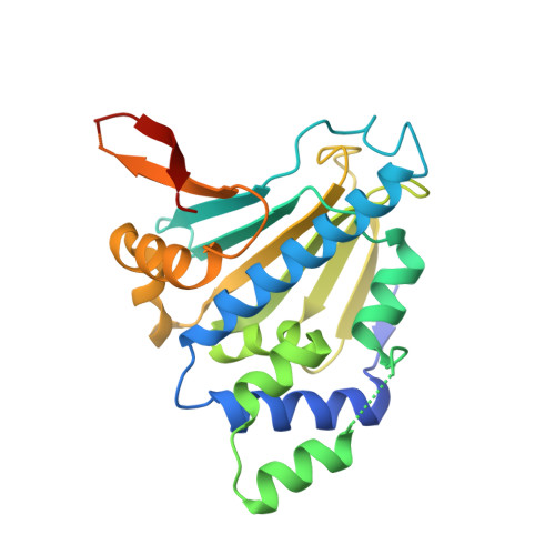
7ULL

Human Grp94 N-terminal domain in complex with 42C


Experimental Data Snapshot

  • Method: X-RAY DIFFRACTION
  • Resolution: 2.31 Å
  • R-Value Free: 0.296 
  • R-Value Work: 0.258 
  • R-Value Observed: 0.261 

Starting Model: experimental
View more details

wwPDB Validation   3D Report Full Report


Ligand Structure Quality Assessment 


This is version 1.2 of the entry. See complete history


Literature

Pan-HSP90 ligand binding reveals isoform-specific differences in plasticity and water networks.

Stachowski, T.R.Nithianantham, S.Vanarotti, M.Lopez, K.Fischer, M.

(2023) Protein Sci 32: e4629-e4629

  • DOI: https://doi.org/10.1002/pro.4629
  • Primary Citation of Related Structures:  
    7ULJ, 7ULK, 7ULL

  • PubMed Abstract: 

    Isoforms of heat shock protein 90 (HSP90) fold oncoproteins that facilitate all 10 hallmarks of cancer. However, its promise as a therapeutic target remains unfulfilled as there is still no FDA-approved drug targeting HSP90 in disease. Among the reasons hindering progress are side effects caused by pan-HSP90 inhibition. Selective targeting of the four isoforms is challenging due to high sequence and structural similarity. Surprisingly, while decades of drug discovery efforts have produced almost 400 human HSP90 structures, no single ligand has been structurally characterized across all four human isoforms to date, which could reveal structural differences to achieve selectivity. To better understand the HSP90 landscape relevant for ligand binding and design we take a three-pronged approach. First, we solved the first complete set of structures of a single ligand bound to all four human isoforms. This enabled a systematic comparison of how side-chains and water networks respond to ligand binding across isoforms. Second, we expanded our analysis to publicly available, incomplete isoform-ligand series with distinct ligand chemistry. This highlighted general trends of protein and water mobility that differ among isoforms and impact ligand binding. Third, we further probed the Hsp90α conformational landscape for accommodating a congeneric series containing the purine scaffold common to HSP90 inhibitors. This revealed how minor ligand modifications flip ligand poses and perturb water and protein conformations. Taken together, this work illustrates how a systematic approach can shed new light on an "old" target and reveal hidden isoform-specific accommodations of congeneric ligands that may be exploited in ligand discovery and design.


  • Organizational Affiliation

    Department of Chemical Biology and Therapeutics, St. Jude Children's Research Hospital, Memphis, Tennessee, USA.


Macromolecules
Find similar proteins by:  (by identity cutoff)  |  3D Structure
Entity ID: 1
MoleculeChains Sequence LengthOrganismDetailsImage
Endoplasmin
A, B
272Homo sapiensMutation(s): 0 
Gene Names: HSP90B1GRP94TRA1
UniProt & NIH Common Fund Data Resources
Find proteins for P14625 (Homo sapiens)
Explore P14625 
Go to UniProtKB:  P14625
PHAROS:  P14625
GTEx:  ENSG00000166598 
Entity Groups  
Sequence Clusters30% Identity50% Identity70% Identity90% Identity95% Identity100% Identity
UniProt GroupP14625
Sequence Annotations
Expand
  • Reference Sequence
Small Molecules
Ligands 4 Unique
IDChains Name / Formula / InChI Key2D Diagram3D Interactions
PG4
Query on PG4

Download Ideal Coordinates CCD File 
D [auth A]TETRAETHYLENE GLYCOL
C8 H18 O5
UWHCKJMYHZGTIT-UHFFFAOYSA-N
42C (Subject of Investigation/LOI)
Query on 42C

Download Ideal Coordinates CCD File 
C [auth A],
G [auth B]
N,N-dimethyl-7H-purin-6-amine
C7 H9 N5
BVIAOQMSVZHOJM-UHFFFAOYSA-N
GOL
Query on GOL

Download Ideal Coordinates CCD File 
F [auth A],
H [auth B]
GLYCEROL
C3 H8 O3
PEDCQBHIVMGVHV-UHFFFAOYSA-N
DMS
Query on DMS

Download Ideal Coordinates CCD File 
E [auth A]DIMETHYL SULFOXIDE
C2 H6 O S
IAZDPXIOMUYVGZ-UHFFFAOYSA-N
Experimental Data & Validation

Experimental Data

  • Method: X-RAY DIFFRACTION
  • Resolution: 2.31 Å
  • R-Value Free: 0.296 
  • R-Value Work: 0.258 
  • R-Value Observed: 0.261 
  • Space Group: P 21 21 2
Unit Cell:
Length ( Å )Angle ( ˚ )
a = 63.921α = 90
b = 89.461β = 90
c = 98.85γ = 90
Software Package:
Software NamePurpose
XDSdata reduction
Aimlessdata scaling
PHENIXrefinement
PDB_EXTRACTdata extraction
PHASERphasing

Structure Validation

View Full Validation Report


Ligand Structure Quality Assessment 


Entry History & Funding Information

Deposition Data


Funding OrganizationLocationGrant Number
National Institutes of Health/National Institute of General Medical Sciences (NIH/NIGMS)United StatesGM142772

Revision History  (Full details and data files)

  • Version 1.0: 2023-04-12
    Type: Initial release
  • Version 1.1: 2023-05-10
    Changes: Database references
  • Version 1.2: 2023-10-25
    Changes: Data collection, Refinement description