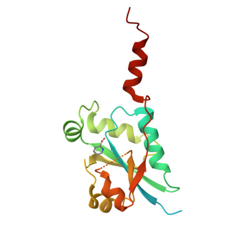
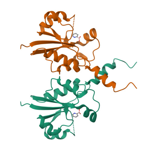
Crystallographic screening using ultra-low-molecular-weight ligands to guide drug design of PfCCT inhibitors.
Duclovel, C., Gelin, M., Wein, S., Wengelnik, K., Krimm, I., Guichou, J.F., Cerdan, R.To be published.
Experimental Data Snapshot
Starting Model: experimental
View more details
Entity ID: 1 | |||||
|---|---|---|---|---|---|
| Molecule | Chains | Sequence Length | Organism | Details | Image |
| Cholinephosphate cytidylyltransferase | 180 | Plasmodium falciparum | Mutation(s): 0 Gene Names: ctP, MAL13P1.86 EC: 2.7.7.15 | 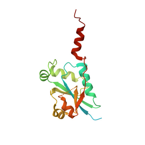 | |
UniProt | |||||
Find proteins for Q8IEE9 (Plasmodium falciparum (isolate 3D7)) Explore Q8IEE9 Go to UniProtKB: Q8IEE9 | |||||
Entity Groups | |||||
| Sequence Clusters | 30% Identity50% Identity70% Identity90% Identity95% Identity100% Identity | ||||
| UniProt Group | Q8IEE9 | ||||
Sequence AnnotationsExpand | |||||
| |||||
| Ligands 1 Unique | |||||
|---|---|---|---|---|---|
| ID | Chains | Name / Formula / InChI Key | 2D Diagram | 3D Interactions | |
| 86Z (Subject of Investigation/LOI) Query on 86Z | B [auth A] | pyridin-2-ylboronic acid C5 H6 B N O2 UMLDUMMLRZFROX-UHFFFAOYSA-N |  | ||
| Length ( Å ) | Angle ( ˚ ) |
|---|---|
| a = 50.401 | α = 90 |
| b = 69.078 | β = 90 |
| c = 117.835 | γ = 90 |
| Software Name | Purpose |
|---|---|
| Aimless | data scaling |
| PHENIX | refinement |
| PDB_EXTRACT | data extraction |
| XDS | data reduction |
| PHASER | phasing |
| Funding Organization | Location | Grant Number |
|---|---|---|
| French Infrastructure for Integrated Structural Biology (FRISBI) | France | -- |
| Montpellier University of Excellence (MUSE) | France | -- |