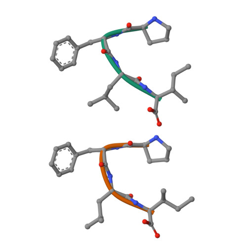
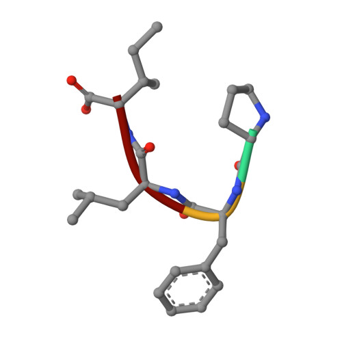
Crystal structure of Pro-Phe-Leu-Ile
Kurumida, Y., Ikeda, K., Nakamichi, Y., Hirano, A., Kobayashi, K., Saito, Y., Kameda, T.To be published.
Experimental Data Snapshot
wwPDB Validation 3D Report Full Report

Find similar proteins by: Sequence | 3D Structure
Entity ID: 1 | |||||
|---|---|---|---|---|---|
| Molecule | Chains | Sequence Length | Organism | Details | Image |
| PRO-PHE-LEU-ILE | 4 | synthetic construct | Mutation(s): 0 | 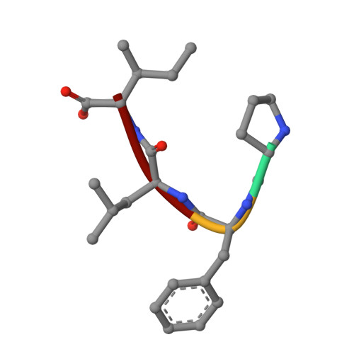 | |
Sequence AnnotationsExpand | |||||
| |||||
| Length ( Å ) | Angle ( ˚ ) |
|---|---|
| a = 5.264 | α = 90 |
| b = 24.967 | β = 94.85 |
| c = 20.736 | γ = 90 |
| Software Name | Purpose |
|---|---|
| SHELX | refinement |
| XDS | data reduction |
| XSCALE | data scaling |
| PDB_EXTRACT | data extraction |
| SHELXT | phasing |
RCSB PDB is hosted by

RCSB PDB is a member of the