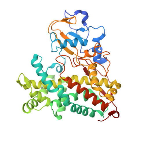
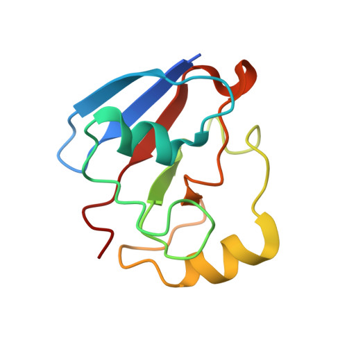
6NBL

Cytochrome P450cam-putidaredoxin complex bound to camphor and cyanide


Experimental Data Snapshot

  • Method: X-RAY DIFFRACTION
  • Resolution: 2.15 Å
  • R-Value Free: 0.254 
  • R-Value Work: 0.195 
  • R-Value Observed: 0.198 

Starting Model: experimental
View more details

wwPDB Validation   3D Report Full Report


Ligand Structure Quality Assessment 


This is version 2.0 of the entry. See complete history


Literature

Ligand and Redox Partner Binding Generates a New Conformational State in Cytochrome P450cam (CYP101A1).

Follmer, A.H.Tripathi, S.Poulos, T.L.

(2019) J Am Chem Soc 141: 2678-2683

  • DOI: https://doi.org/10.1021/jacs.8b13079
  • Primary Citation of Related Structures:  
    6NBL

  • PubMed Abstract: 

    It has become increasingly clear that cytochromes P450 can cycle back and forth between two extreme conformational states termed the closed and open states. In the well-studied cytochrome P450cam, the binding of its redox partner, putidaredoxin (Pdx), shifts P450cam toward the open state. Shifting to the open state is thought to be important in the formation of a proton relay network essential for O-O bond cleavage and formation of the active Fe(IV)═O intermediate. Another important intermediate is the oxy-P450cam complex when bound to Pdx. Trapping this intermediate in crystallo is challenging owing to its instability, but the CN - complex is both stable and an excellent mimic of the O 2 complex. Here we present the P450cam-Pdx structure complexed with CN - . CN - results in large conformational changes including cis/trans isomerization of proline residues. Changes include large rearrangements of active-site residues and the formation of new active-site access channel that we have termed channel 2. The formation of channel 2 has also been observed in our previous molecular dynamics simulations wherein substrate binding to an allosteric site remote from the active site opens up channel 2. This new structure supports an extensive amount of previous work showing that distant regions of the structure are dynamically coupled and underscores the potentially important role that large conformational changes and dynamics play in P450 catalysis.


  • Organizational Affiliation

    Departments of Molecular Biology and Biochemistry, Pharmaceutical Sciences, and Chemistry , University of California , Irvine , California 92697-3900 , United States.


Macromolecules
Find similar proteins by:  (by identity cutoff)  |  3D Structure
Entity ID: 1
MoleculeChains Sequence LengthOrganismDetailsImage
Camphor 5-monooxygenase
A, B
415Pseudomonas putidaMutation(s): 6 
Gene Names: camCcyp101
EC: 1.14.15.1
UniProt
Find proteins for P00183 (Pseudomonas putida)
Explore P00183 
Go to UniProtKB:  P00183
Entity Groups  
Sequence Clusters30% Identity50% Identity70% Identity90% Identity95% Identity100% Identity
UniProt GroupP00183
Sequence Annotations
Expand
  • Reference Sequence
Find similar proteins by:  (by identity cutoff)  |  3D Structure
Entity ID: 2
MoleculeChains Sequence LengthOrganismDetailsImage
Putidaredoxin
C, D
112Pseudomonas putidaMutation(s): 2 
Gene Names: camB
UniProt
Find proteins for P00259 (Pseudomonas putida)
Explore P00259 
Go to UniProtKB:  P00259
Entity Groups  
Sequence Clusters30% Identity50% Identity70% Identity90% Identity95% Identity100% Identity
UniProt GroupP00259
Sequence Annotations
Expand
  • Reference Sequence
Small Molecules
Ligands 6 Unique
IDChains Name / Formula / InChI Key2D Diagram3D Interactions
HEM
Query on HEM

Download Ideal Coordinates CCD File 
E [auth A],
I [auth B]
PROTOPORPHYRIN IX CONTAINING FE
C34 H32 Fe N4 O4
KABFMIBPWCXCRK-RGGAHWMASA-L
1N0
Query on 1N0

Download Ideal Coordinates CCD File 
M [auth B],
O [auth C]
1,1'-hexane-1,6-diyldipyrrolidine-2,5-dione
C14 H20 N2 O4
PBFKSBAPGGMKKJ-UHFFFAOYSA-N
FES
Query on FES

Download Ideal Coordinates CCD File 
N [auth C],
P [auth D]
FE2/S2 (INORGANIC) CLUSTER
Fe2 S2
NIXDOXVAJZFRNF-UHFFFAOYSA-N
CAM
Query on CAM

Download Ideal Coordinates CCD File 
G [auth A],
K [auth B]
CAMPHOR
C10 H16 O
DSSYKIVIOFKYAU-XCBNKYQSSA-N
CA
Query on CA

Download Ideal Coordinates CCD File 
H [auth A],
L [auth B]
CALCIUM ION
Ca
BHPQYMZQTOCNFJ-UHFFFAOYSA-N
CYN
Query on CYN

Download Ideal Coordinates CCD File 
F [auth A],
J [auth B]
CYANIDE ION
C N
XFXPMWWXUTWYJX-UHFFFAOYSA-N
Experimental Data & Validation

Experimental Data

  • Method: X-RAY DIFFRACTION
  • Resolution: 2.15 Å
  • R-Value Free: 0.254 
  • R-Value Work: 0.195 
  • R-Value Observed: 0.198 
  • Space Group: P 1 21 1
Unit Cell:
Length ( Å )Angle ( ˚ )
a = 57.36α = 90
b = 110.3β = 107.67
c = 88.62γ = 90
Software Package:
Software NamePurpose
PHENIXrefinement
MOSFLMdata reduction
SCALAdata scaling
PHASERphasing

Structure Validation

View Full Validation Report


Ligand Structure Quality Assessment 


Entry History & Funding Information

Deposition Data


Funding OrganizationLocationGrant Number
National Institutes of Health/National Institute of General Medical Sciences (NIH/NIGMS)United StatesGM057353

Revision History  (Full details and data files)

  • Version 1.0: 2019-03-06
    Type: Initial release
  • Version 1.1: 2020-01-01
    Changes: Author supporting evidence
  • Version 1.2: 2023-10-11
    Changes: Data collection, Database references, Derived calculations, Refinement description
  • Version 2.0: 2024-12-25
    Changes: Advisory, Atomic model, Data collection, Derived calculations, Structure summary