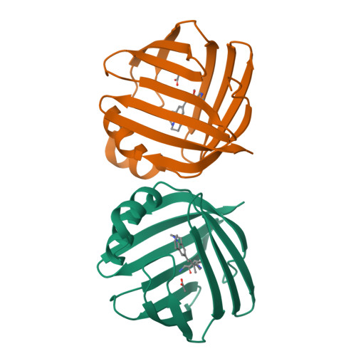
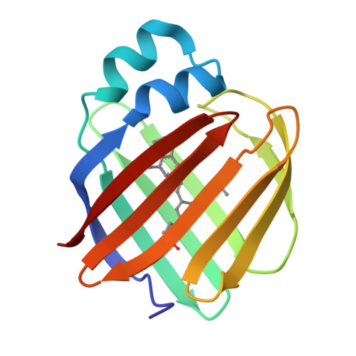
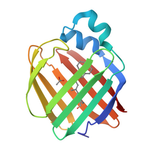
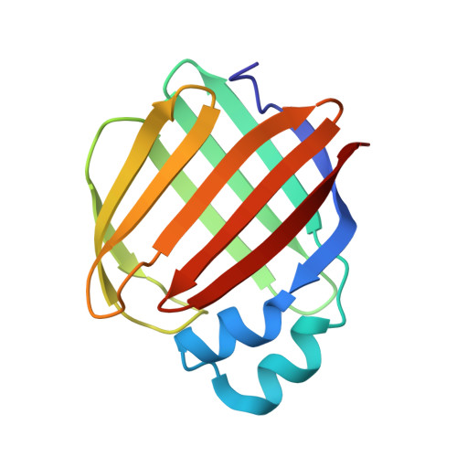
6C7Z | pdb_00006c7z

Crystal structure of the Q108K:K40L:T51V:R58F mutant of human Cellular Retinol Binding Protein II in complex with synthetic Ligand Julolidine


Experimental Data Snapshot

  • Method: X-RAY DIFFRACTION
  • Resolution: 1.42 Å
  • R-Value Free: 
    0.256 (Depositor), 0.260 (DCC) 
  • R-Value Work: 
    0.222 (Depositor), 0.220 (DCC) 
  • R-Value Observed: 
    0.224 (Depositor) 

Starting Model: experimental
View more details

wwPDB Validation   3D Report Full Report


Ligand Structure Quality Assessment 

Created with Raphaël 2.3.0Worse 01 BetterLigand structure goodness of fit to experimental dataBest fitted JLOClick on this verticalbar to view details

This is version 1.5 of the entry. See complete history


Literature

A Genetically Encoded Ratiometric pH Probe: Wavelength Regulation-Inspired Design of pH Indicators.

Berbasova, T.Tahmasebi Nick, S.Nosrati, M.Nossoni, Z.Santos, E.M.Vasileiou, C.Geiger, J.H.Borhan, B.

(2018) Chembiochem 19: 1288-1295

  • DOI: https://doi.org/10.1002/cbic.201800050
  • Primary Citation of Related Structures:  
    6C7Z

  • PubMed Abstract: 

    Mutants of human cellular retinol-binding protein II (hCRBPII) were engineered to bind a julolidine retinal analogue for the purpose of developing a ratiometric pH sensor. The design relied on the electrostatic influence of a titratable amino acid side chain, which affects the absorption and, thus, the emission of the protein/fluorophore complex. The ratio of emissions obtained at two excitation wavelengths that correspond to the absorption of the two forms of the protein/fluorophore complex, leads to a concentration-independent measure of pH.


  • Organizational Affiliation

    Department of Chemistry, Michigan State University, East Lansing, MI, 48824, USA.


Macromolecules
Find similar proteins by:  (by identity cutoff)  |  3D Structure
Entity ID: 1
MoleculeChains Sequence LengthOrganismDetailsImage
Retinol-binding protein 2
A, B
133Homo sapiensMutation(s): 4 
Gene Names: RBP2CRBP2
UniProt & NIH Common Fund Data Resources
Find proteins for P50120 (Homo sapiens)
Explore P50120 
Go to UniProtKB:  P50120
PHAROS:  P50120
GTEx:  ENSG00000114113 
Entity Groups  
Sequence Clusters30% Identity50% Identity70% Identity90% Identity95% Identity100% Identity
UniProt GroupP50120
Sequence Annotations
Expand
  • Reference Sequence
Experimental Data & Validation

Experimental Data

  • Method: X-RAY DIFFRACTION
  • Resolution: 1.42 Å
  • R-Value Free:  0.256 (Depositor), 0.260 (DCC) 
  • R-Value Work:  0.222 (Depositor), 0.220 (DCC) 
  • R-Value Observed: 0.224 (Depositor) 
Space Group: P 1
Unit Cell:
Length ( Å )Angle ( ˚ )
a = 29.787α = 90.51
b = 35.947β = 92.45
c = 64.089γ = 113.31
Software Package:
Software NamePurpose
PHENIXrefinement
HKL-3000data reduction
HKL-3000data scaling
PHENIXphasing

Structure Validation

View Full Validation Report


Ligand Structure Quality Assessment 

Created with Raphaël 2.3.0Worse 01 BetterLigand structure goodness of fit to experimental dataBest fitted JLOClick on this verticalbar to view details

Entry History & Funding Information

Deposition Data


Funding OrganizationLocationGrant Number
National Institutes of Health/National Institute of General Medical Sciences (NIH/NIGMS)United StatesGM101353

Revision History  (Full details and data files)

  • Version 1.0: 2018-04-25
    Type: Initial release
  • Version 1.1: 2018-07-04
    Changes: Data collection, Database references
  • Version 1.2: 2019-02-20
    Changes: Author supporting evidence, Data collection
  • Version 1.3: 2022-03-23
    Changes: Author supporting evidence, Database references
  • Version 1.4: 2023-10-04
    Changes: Data collection, Refinement description
  • Version 1.5: 2024-11-13
    Changes: Structure summary