5ZE9

Crystal structure of AMP-PNP bound mutant A3B3 complex from Enterococcus hirae V-ATPase


Experimental Data Snapshot

  • Method: X-RAY DIFFRACTION
  • Resolution: 2.10 Å
  • R-Value Free: 
    0.196 (Depositor), 0.200 (DCC) 
  • R-Value Work: 
    0.166 (Depositor), 0.170 (DCC) 
  • R-Value Observed: 
    0.168 (Depositor) 

Starting Model: experimental
View more details

wwPDB Validation   3D Report Full Report


Ligand Structure Quality Assessment 

Created with Raphaël 2.3.0Worse 01 BetterLigand structure goodness of fit to experimental dataBest fitted ANPClick on this verticalbar to view details

This is version 1.2 of the entry. See complete history


Literature

Metastable asymmetrical structure of a shaftless V1motor.

Maruyama, S.Suzuki, K.Imamura, M.Sasaki, H.Matsunami, H.Mizutani, K.Saito, Y.Imai, F.L.Ishizuka-Katsura, Y.Kimura-Someya, T.Shirouzu, M.Uchihashi, T.Ando, T.Yamato, I.Murata, T.

(2019) Sci Adv 5: eaau8149-eaau8149

  • DOI: https://doi.org/10.1126/sciadv.aau8149
  • Primary Citation of Related Structures:  
    5ZE9, 5ZEA

  • PubMed Abstract: 

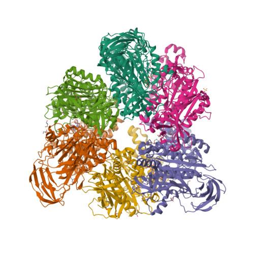
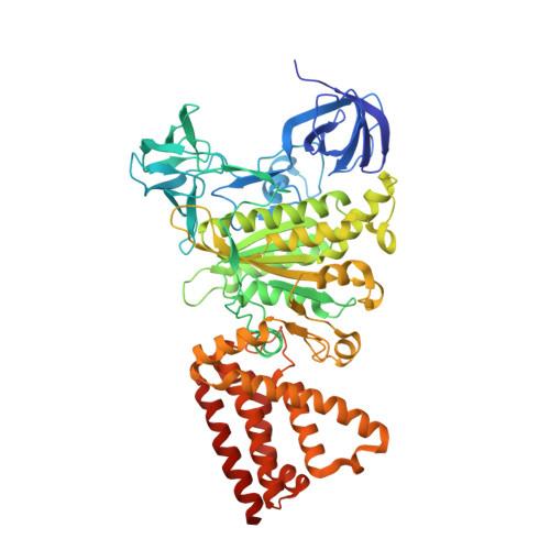
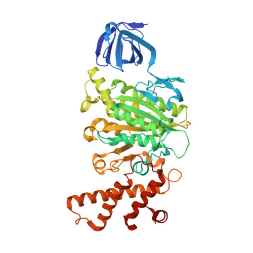
    V 1 -ATPase is an ATP-driven rotary motor that is composed of a ring-shaped A 3 B 3 complex and a central DF shaft. The nucleotide-free A 3 B 3 complex of Enterococcus hirae , composed of three identical A 1 B 1 heterodimers, showed a unique asymmetrical structure, probably due to the strong binding of the N-terminal barrel domain, which forms a crown structure. Here, we mutated the barrel region to weaken the crown, and performed structural analyses using high-speed atomic force microscopy and x-ray crystallography of the mutant A 3 B 3 . The nucleotide-free mutant A 3 B 3 complex had a more symmetrical open structure than the wild type. Binding of nucleotides produced a closely packed spiral-like structure with a disrupted crown. These findings suggest that wild-type A 3 B 3 forms a metastable (stressed) asymmetric structure composed of unstable A 1 B 1 conformers due to the strong constraint of the crown. The results further the understanding of the principle of the cooperative transition mechanism of rotary motors.


  • Organizational Affiliation

    Department of Chemistry, Graduate School of Science, Chiba University, 1-33 Yayoi-cho, Inage, Chiba 263-8522, Japan.


Macromolecules
Find similar proteins by:  (by identity cutoff)  |  3D Structure
Entity ID: 1
MoleculeChains Sequence LengthOrganismDetailsImage
V-type sodium ATPase catalytic subunit A
A, B, C
600Enterococcus hirae ATCC 9790Mutation(s): 0 
Gene Names: ntpAEHR_08260
EC: 3.6.3.15 (PDB Primary Data), 7.2.2.1 (UniProt)
UniProt
Find proteins for Q08636 (Enterococcus hirae (strain ATCC 9790 / DSM 20160 / JCM 8729 / LMG 6399 / NBRC 3181 / NCIMB 6459 / NCDO 1258 / NCTC 12367 / WDCM 00089 / R))
Explore Q08636 
Go to UniProtKB:  Q08636
Entity Groups  
Sequence Clusters30% Identity50% Identity70% Identity90% Identity95% Identity100% Identity
UniProt GroupQ08636
Sequence Annotations
Expand
  • Reference Sequence
Find similar proteins by:  (by identity cutoff)  |  3D Structure
Entity ID: 2
MoleculeChains Sequence LengthOrganismDetailsImage
V-type sodium ATPase subunit B
D, E, F
465Enterococcus hirae ATCC 9790Mutation(s): 1 
Gene Names: ntpBEHR_08265
UniProt
Find proteins for Q08637 (Enterococcus hirae (strain ATCC 9790 / DSM 20160 / JCM 8729 / LMG 6399 / NBRC 3181 / NCIMB 6459 / NCDO 1258 / NCTC 12367 / WDCM 00089 / R))
Explore Q08637 
Go to UniProtKB:  Q08637
Entity Groups  
Sequence Clusters30% Identity50% Identity70% Identity90% Identity95% Identity100% Identity
UniProt GroupQ08637
Sequence Annotations
Expand
  • Reference Sequence
Small Molecules
Ligands 4 Unique
IDChains Name / Formula / InChI Key2D Diagram3D Interactions
ANP
Query on ANP

Download Ideal Coordinates CCD File 
BA [auth C],
G [auth A],
P [auth B]
PHOSPHOAMINOPHOSPHONIC ACID-ADENYLATE ESTER
C10 H17 N6 O12 P3
PVKSNHVPLWYQGJ-KQYNXXCUSA-N
MES
Query on MES

Download Ideal Coordinates CCD File 
LA [auth D]2-(N-MORPHOLINO)-ETHANESULFONIC ACID
C6 H13 N O4 S
SXGZJKUKBWWHRA-UHFFFAOYSA-N
GOL
Query on GOL

Download Ideal Coordinates CCD File 
AA [auth B]
DA [auth C]
EA [auth C]
FA [auth C]
GA [auth C]
GLYCEROL
C3 H8 O3
PEDCQBHIVMGVHV-UHFFFAOYSA-N
MG
Query on MG

Download Ideal Coordinates CCD File 
CA [auth C],
H [auth A],
Q [auth B]
MAGNESIUM ION
Mg
JLVVSXFLKOJNIY-UHFFFAOYSA-N
Experimental Data & Validation

Experimental Data

  • Method: X-RAY DIFFRACTION
  • Resolution: 2.10 Å
  • R-Value Free:  0.196 (Depositor), 0.200 (DCC) 
  • R-Value Work:  0.166 (Depositor), 0.170 (DCC) 
  • R-Value Observed: 0.168 (Depositor) 
Space Group: P 21 21 21
Unit Cell:
Length ( Å )Angle ( ˚ )
a = 105.83α = 90
b = 151.37β = 90
c = 235.49γ = 90
Software Package:
Software NamePurpose
XDSdata reduction
HKL-2000data scaling
MOLREPphasing
PHENIXrefinement
PDB_EXTRACTdata extraction

Structure Validation

View Full Validation Report


Ligand Structure Quality Assessment 

Created with Raphaël 2.3.0Worse 01 BetterLigand structure goodness of fit to experimental dataBest fitted ANPClick on this verticalbar to view details

Entry History 

Deposition Data

Revision History  (Full details and data files)

  • Version 1.0: 2019-02-06
    Type: Initial release
  • Version 1.1: 2019-02-20
    Changes: Data collection, Database references
  • Version 1.2: 2023-11-22
    Changes: Data collection, Database references, Derived calculations, Refinement description