5U8L | pdb_00005u8l

Crystal structure of EGFR kinase domain in complex with a sulfonyl fluoride probe XO44


Experimental Data Snapshot

  • Method: X-RAY DIFFRACTION
  • Resolution: 1.60 Å
  • R-Value Free: 
    0.202 (Depositor), 0.200 (DCC) 
  • R-Value Work: 
    0.189 (Depositor), 0.180 (DCC) 
  • R-Value Observed: 
    0.190 (Depositor) 

wwPDB Validation   3D Report Full Report


Ligand Structure Quality Assessment 

Created with Raphaël 2.3.0Worse 01 BetterLigand structure goodness of fit to experimental dataBest fitted O44Click on this verticalbar to view details

This is version 1.2 of the entry. See complete history


Literature

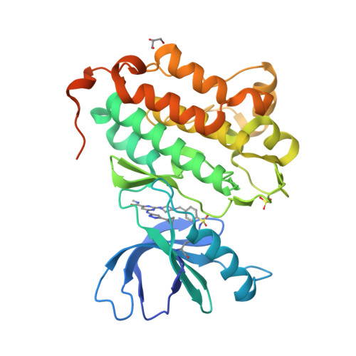
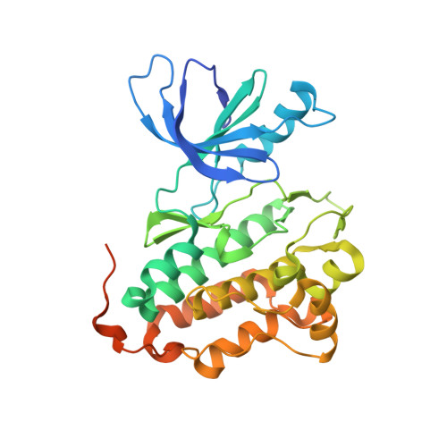
Broad-Spectrum Kinase Profiling in Live Cells with Lysine-Targeted Sulfonyl Fluoride Probes.

Zhao, Q.Ouyang, X.Wan, X.Gajiwala, K.S.Kath, J.C.Jones, L.H.Burlingame, A.L.Taunton, J.

(2017) J Am Chem Soc 139: 680-685

  • DOI: https://doi.org/10.1021/jacs.6b08536
  • Primary Citation of Related Structures:  
    5K9I, 5U8L

  • PubMed Abstract: 

    Protein kinases comprise a large family of structurally related enzymes. A major goal in kinase-inhibitor development is to selectively engage the desired kinase while avoiding myriad off-target kinases. However, quantifying inhibitor interactions with multiple endogenous kinases in live cells remains an unmet challenge. Here, we report the design of sulfonyl fluoride probes that covalently label a broad swath of the intracellular kinome with high efficiency. Protein crystallography and mass spectrometry confirmed a chemoselective reaction between the sulfonyl fluoride and a conserved lysine in the ATP binding site. Optimized probe 2 (XO44) covalently modified up to 133 endogenous kinases, efficiently competing with high intracellular concentrations of ATP. We employed probe 2 and label-free mass spectrometry to quantify intracellular kinase engagement by the approved drug, dasatinib. The data revealed saturable dasatinib binding to a small subset of kinase targets at clinically relevant concentrations, highlighting the utility of lysine-targeted sulfonyl fluoride probes in demanding chemoproteomic applications.


  • Organizational Affiliation

    Worldwide Research and Development, Pfizer , San Diego, California 92121, United States.


Macromolecules
Find similar proteins by:  (by identity cutoff)  |  3D Structure
Entity ID: 1
MoleculeChains Sequence LengthOrganismDetailsImage
Epidermal growth factor receptor329Homo sapiensMutation(s): 0 
Gene Names: EGFRERBBERBB1HER1
EC: 2.7.10.1
UniProt & NIH Common Fund Data Resources
Find proteins for P00533 (Homo sapiens)
Explore P00533 
Go to UniProtKB:  P00533
PHAROS:  P00533
GTEx:  ENSG00000146648 
Entity Groups  
Sequence Clusters30% Identity50% Identity70% Identity90% Identity95% Identity100% Identity
UniProt GroupP00533
Sequence Annotations
Expand
  • Reference Sequence
Small Molecules
Ligands 3 Unique
IDChains Name / Formula / InChI Key2D Diagram3D Interactions
O44
Query on O44

Download Ideal Coordinates CCD File 
B [auth A]4-[(4-{4-[(3-cyclopropyl-1H-pyrazol-5-yl)amino]-6-[(prop-2-yn-1-yl)carbamoyl]pyrimidin-2-yl}piperazin-1-yl)methyl]benzene-1-sulfonyl fluoride
C25 H27 F N8 O3 S
ZBSPMOBILDLOCV-UHFFFAOYSA-N
SO4
Query on SO4

Download Ideal Coordinates CCD File 
C [auth A]SULFATE ION
O4 S
QAOWNCQODCNURD-UHFFFAOYSA-L
GOL
Query on GOL

Download Ideal Coordinates CCD File 
D [auth A]GLYCEROL
C3 H8 O3
PEDCQBHIVMGVHV-UHFFFAOYSA-N
Experimental Data & Validation

Experimental Data

  • Method: X-RAY DIFFRACTION
  • Resolution: 1.60 Å
  • R-Value Free:  0.202 (Depositor), 0.200 (DCC) 
  • R-Value Work:  0.189 (Depositor), 0.180 (DCC) 
  • R-Value Observed: 0.190 (Depositor) 
Space Group: P 21 21 21
Unit Cell:
Length ( Å )Angle ( ˚ )
a = 40.364α = 90
b = 69.977β = 90
c = 110.403γ = 90
Software Package:
Software NamePurpose
CNXrefinement
XDSdata reduction
Aimlessdata scaling
CNXphasing

Structure Validation

View Full Validation Report


Ligand Structure Quality Assessment 

Created with Raphaël 2.3.0Worse 01 BetterLigand structure goodness of fit to experimental dataBest fitted O44Click on this verticalbar to view details

Entry History 

Deposition Data

Revision History  (Full details and data files)

  • Version 1.0: 2017-01-25
    Type: Initial release
  • Version 1.1: 2017-02-01
    Changes: Database references
  • Version 1.2: 2024-10-30
    Changes: Data collection, Database references, Structure summary