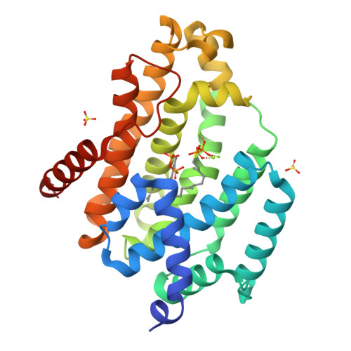
Crystal structure of a dehydrosqualene synthase in complex with ligand
Liu, G.Z., Liu, W.D., Chen, C.C., Guo, R.T.To be published.
Experimental Data Snapshot
Starting Model: experimental
View more details
Entity ID: 1 | |||||
|---|---|---|---|---|---|
| Molecule | Chains | Sequence Length | Organism | Details | Image |
| Phytoene synthase | 289 | Enterococcus hirae ATCC 9790 | Mutation(s): 0 |  | |
UniProt | |||||
Find proteins for I6T9U8 (Enterococcus hirae (strain ATCC 9790 / DSM 20160 / JCM 8729 / LMG 6399 / NBRC 3181 / NCIMB 6459 / NCDO 1258 / NCTC 12367 / WDCM 00089 / R)) Explore I6T9U8 Go to UniProtKB: I6T9U8 | |||||
Entity Groups | |||||
| Sequence Clusters | 30% Identity50% Identity70% Identity90% Identity95% Identity100% Identity | ||||
| UniProt Group | I6T9U8 | ||||
Sequence AnnotationsExpand | |||||
| |||||
| Ligands 3 Unique | |||||
|---|---|---|---|---|---|
| ID | Chains | Name / Formula / InChI Key | 2D Diagram | 3D Interactions | |
| FPS Query on FPS | G [auth A], H [auth A] | S-[(2E,6E)-3,7,11-TRIMETHYLDODECA-2,6,10-TRIENYL] TRIHYDROGEN THIODIPHOSPHATE C15 H28 O6 P2 S MYMLCRQRXFRQGP-YFVJMOTDSA-N |  | ||
| SO4 Query on SO4 | B [auth A], C [auth A] | SULFATE ION O4 S QAOWNCQODCNURD-UHFFFAOYSA-L |  | ||
| MG Query on MG | D [auth A], E [auth A], F [auth A] | MAGNESIUM ION Mg JLVVSXFLKOJNIY-UHFFFAOYSA-N |  | ||
| Length ( Å ) | Angle ( ˚ ) |
|---|---|
| a = 37.238 | α = 90 |
| b = 41.09 | β = 96.35 |
| c = 93.477 | γ = 90 |
| Software Name | Purpose |
|---|---|
| HKL-2000 | data collection |
| HKL-2000 | data scaling |
| PHASER | phasing |
| REFMAC | refinement |
| PDB_EXTRACT | data extraction |
| HKL-2000 | data reduction |
| PHASER | phasing |
RCSB PDB is hosted by

RCSB PDB is a member of the