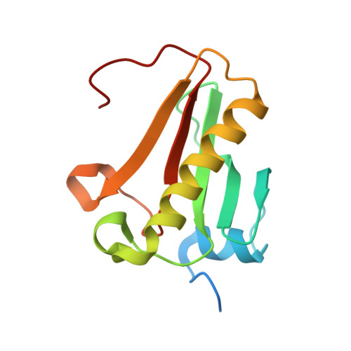
Caught before Released: Structural Mapping of the Reaction Trajectory for the Sofosbuvir Activating Enzyme, Human Histidine Triad Nucleotide Binding Protein 1 (hHint1).
Shah, R., Maize, K.M., Zhou, X., Finzel, B.C., Wagner, C.R.(2017) Biochemistry 56: 3559-3570
- PubMed: 28691797
- DOI: https://doi.org/10.1021/acs.biochem.7b00148
- Primary Citation of Related Structures:
5IPB, 5IPC, 5IPD, 5IPE - PubMed Abstract:
Human histidine triad nucleotide binding protein 1 (hHint1) is classified as an efficient nucleoside phosphoramidase and acyl-adenosine monophosphate hydrolase. Human Hint1 has been shown to be essential for the metabolic activation of nucleotide antiviral pronucleotides (i.e., proTides), such as the FDA approved hepatitis C drug, sofosbuvir. The active site of hHint1 comprises an ensemble of strictly conserved histidines, including nucleophilic His112. To structurally investigate the mechanism of hHint1 catalysis, we have designed and prepared nucleoside thiophosphoramidate substrates that are able to capture the transiently formed nucleotidylated-His112 intermediate (E*) using time-dependent crystallography. Utilizing a catalytically inactive hHint1 His112Asn enzyme variant and wild-type enzyme, the enzyme-substrate (ES 1 ) and product (EP 2 ) complexes were also cocrystallized, respectively, thus providing a structural map of the reaction trajectory. On the basis of these observations and the mechanistic necessity of proton transfers, proton inventory studies were carried out. Although we cannot completely exclude the possibility of more than one proton in flight, the results of these studies were consistent with the transfer of a single proton during the formation of the intermediate. Interestingly, structural analysis revealed that the critical proton transfers required for intermediate formation and hydrolysis may be mediated by a conserved active site water channel. Taken together, our results provide mechanistic insights underpinning histidine nucleophilic catalysis in general and hHint1 catalysis, in particular, thus aiding the design of future proTides and the elucidation of the natural function of the Hint family of enzymes.
Organizational Affiliation:
Department of Medicinal Chemistry University of Minnesota , Minneapolis, Minnesota 55455, United States.


