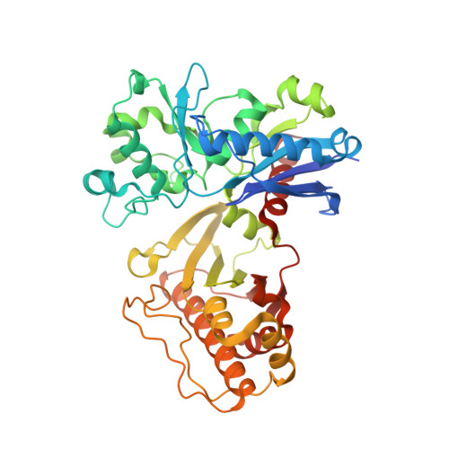
Crystal Structures of Putative Sugar Kinases from Synechococcus Elongatus PCC 7942 and Arabidopsis Thaliana
Xie, Y., Li, M., Chang, W.(2016) PLoS One 11: e0156067-e0156067
- PubMed: 27223615
- DOI: https://doi.org/10.1371/journal.pone.0156067
- Primary Citation of Related Structures:
5HTJ, 5HTN, 5HTP, 5HTR, 5HTV, 5HTX, 5HTY, 5HU2, 5HUX, 5HV7 - PubMed Abstract:
The genome of the Synechococcus elongatus strain PCC 7942 encodes a putative sugar kinase (SePSK), which shares 44.9% sequence identity with the xylulose kinase-1 (AtXK-1) from Arabidopsis thaliana. Sequence alignment suggests that both kinases belong to the ribulokinase-like carbohydrate kinases, a sub-family of FGGY family carbohydrate kinases. However, their exact physiological function and real substrates remain unknown. Here we solved the structures of SePSK and AtXK-1 in both their apo forms and in complex with nucleotide substrates. The two kinases exhibit nearly identical overall architecture, with both kinases possessing ATP hydrolysis activity in the absence of substrates. In addition, our enzymatic assays suggested that SePSK has the capability to phosphorylate D-ribulose. In order to understand the catalytic mechanism of SePSK, we solved the structure of SePSK in complex with D-ribulose and found two potential substrate binding pockets in SePSK. Using mutation and activity analysis, we further verified the key residues important for its catalytic activity. Moreover, our structural comparison with other family members suggests that there are major conformational changes in SePSK upon substrate binding, facilitating the catalytic process. Together, these results provide important information for a more detailed understanding of the cofactor and substrate binding mode as well as the catalytic mechanism of SePSK, and possible similarities with its plant homologue AtXK-1.
Organizational Affiliation:
National Laboratory of Biomacromolecules, CAS Center for Excellence in Biomacromolecules, Institute of Biophysics, Chinese Academy of Sciences, Beijing, China.


