4QYK | pdb_00004qyk

Crystal structure of a canine parvovirus variant


Experimental Data Snapshot

  • Method: X-RAY DIFFRACTION
  • Resolution: 3.50 Å
  • R-Value Free: 
    0.221 (Depositor), 0.222 (DCC) 
  • R-Value Work: 
    0.180 (Depositor), 0.183 (DCC) 
  • R-Value Observed: 
    0.182 (Depositor) 

wwPDB Validation   3D Report Full Report


This is version 1.4 of the entry. See complete history


Literature

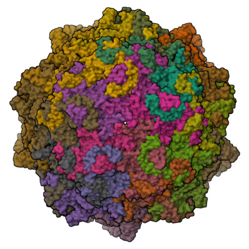
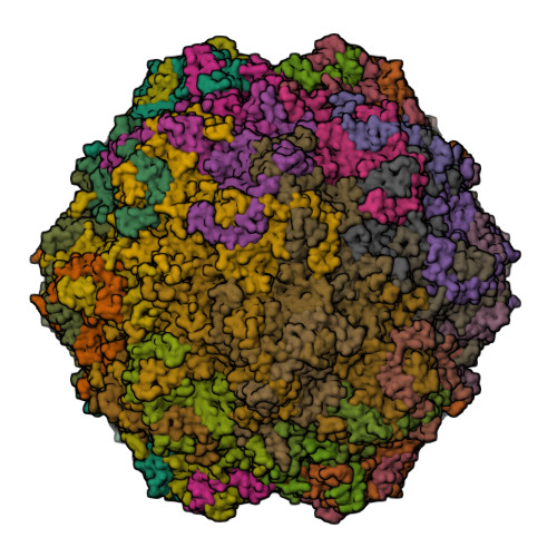
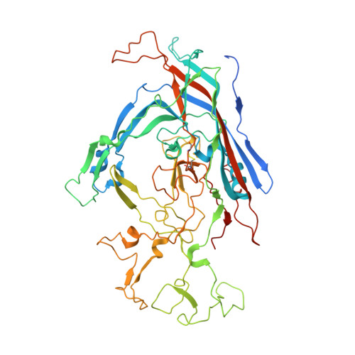
Global Displacement of Canine Parvovirus by a Host-Adapted Variant: Structural Comparison between Pandemic Viruses with Distinct Host Ranges.

Organtini, L.J.Allison, A.B.Lukk, T.Parrish, C.R.Hafenstein, S.

(2015) J Virol 89: 1909-1912

  • DOI: https://doi.org/10.1128/JVI.02611-14
  • Primary Citation of Related Structures:  
    4QYK

  • PubMed Abstract: 

    Canine parvovirus type 2 (CPV-2) emerged in 1978 and spread worldwide within 2 years. Subsequently, CPV-2 was completely replaced by the variant CPV-2a, which is characterized by four specific capsid (VP2) mutations. The X-ray crystal structure of the CPV-2a capsid shows that each mutation confers small local changes. The loss of a hydrogen bond and introduction of a glycine residue likely introduce flexibility to sites that control interactions with the host receptor, antibodies, and sialic acids.


  • Organizational Affiliation

    Department of Medicine, The Pennsylvania State University College of Medicine, Hershey, Pennsylvania, USA.


Macromolecules
Find similar proteins by:  (by identity cutoff)  |  3D Structure
Entity ID: 1
MoleculeChains Sequence LengthOrganismDetailsImage
Capsid protein VP1584Feline panleukopenia virusMutation(s): 0 
UniProt
Find proteins for P90456 (Feline panleukopenia virus)
Explore P90456 
Go to UniProtKB:  P90456
Entity Groups  
Sequence Clusters30% Identity50% Identity70% Identity90% Identity95% Identity100% Identity
UniProt GroupP90456
Sequence Annotations
Expand
  • Reference Sequence
Small Molecules
Ligands 1 Unique
IDChains Name / Formula / InChI Key2D Diagram3D Interactions
MG
Query on MG

Download Ideal Coordinates CCD File 
AB [auth W]
BB [auth X]
CB [auth Y]
DB [auth Y]
EA [auth A]
MAGNESIUM ION
Mg
JLVVSXFLKOJNIY-UHFFFAOYSA-N
Experimental Data & Validation

Experimental Data

  • Method: X-RAY DIFFRACTION
  • Resolution: 3.50 Å
  • R-Value Free:  0.221 (Depositor), 0.222 (DCC) 
  • R-Value Work:  0.180 (Depositor), 0.183 (DCC) 
  • R-Value Observed: 0.182 (Depositor) 
Space Group: P 42 21 2
Unit Cell:
Length ( Å )Angle ( ˚ )
a = 453.1α = 90
b = 453.1β = 90
c = 319.02γ = 90
Software Package:
Software NamePurpose
Aimlessdata scaling
PHENIXrefinement
PDB_EXTRACTdata extraction
HKL-2000data collection
MOSFLMdata reduction
PHASERphasing

Structure Validation

View Full Validation Report


Entry History 

Deposition Data

Revision History  (Full details and data files)

  • Version 1.0: 2014-12-10
    Type: Initial release
  • Version 1.1: 2014-12-17
    Changes: Advisory
  • Version 1.2: 2014-12-24
    Changes: Structure summary
  • Version 1.3: 2015-01-28
    Changes: Database references
  • Version 1.4: 2024-10-30
    Changes: Data collection, Database references, Derived calculations, Structure summary