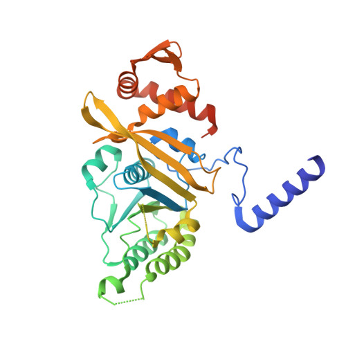
4PTL

Mycobacterium tuberculosis RecA glycerol bound low temperature structure IIC-GM


Experimental Data Snapshot

  • Method: X-RAY DIFFRACTION
  • Resolution: 2.50 Å
  • R-Value Free: 0.248 
  • R-Value Work: 0.201 
  • R-Value Observed: 0.205 

Starting Model: experimental
View more details

wwPDB Validation   3D Report Full Report


This is version 1.2 of the entry. See complete history


Literature

Structural studies on Mycobacterium tuberculosis RecA: Molecular plasticity and interspecies variability

Chandran, A.V.Prabu, J.R.Nautiyal, A.Patil, K.N.Muniyappa, K.Vijayan, M.

(2015) J Biosci 40: 13-30

  • DOI: https://doi.org/10.1007/s12038-014-9497-x
  • Primary Citation of Related Structures:  
    4OQF, 4PO1, 4PO8, 4PO9, 4POA, 4PPF, 4PPG, 4PPN, 4PPQ, 4PQF, 4PQR, 4PQY, 4PR0, 4PSA, 4PSK, 4PSV, 4PTL

  • PubMed Abstract: 

    Structures of crystals of Mycobacterium tuberculosis RecA, grown and analysed under different conditions, provide insights into hitherto underappreciated details of molecular structure and plasticity. In particular, they yield information on the invariant and variable features of the geometry of the P-loop, whose binding to ATP is central for all the biochemical activities of RecA. The strengths of interaction of the ligands with the P-loop reveal significant differences. This in turn affects the magnitude of the motion of the 'switch' residue, Gln195 in M. tuberculosis RecA, which triggers the transmission of ATP-mediated allosteric information to the DNA binding region. M. tuberculosis RecA is substantially rigid compared with its counterparts from M. smegmatis and E. coli, which exhibit concerted internal molecular mobility. The interspecies variability in the plasticity of the two mycobacterial proteins is particularly surprising as they have similar sequence and 3D structure. Details of the interactions of ligands with the protein, characterized in the structures reported here, could be useful for design of inhibitors against M. tuberculosis RecA.


  • Organizational Affiliation

    Molecular Biophysics Unit, Indian Institute of Science, Bangalore 560 012.


Macromolecules
Find similar proteins by:  (by identity cutoff)  |  3D Structure
Entity ID: 1
MoleculeChains Sequence LengthOrganismDetailsImage
Protein RecA, 1st part, 2nd part350Mycobacterium tuberculosisMutation(s): 0 
Gene Names: MT2806MTV002.02crecARv2737c
EC: 3.1
UniProt
Find proteins for P9WHJ3 (Mycobacterium tuberculosis (strain ATCC 25618 / H37Rv))
Explore P9WHJ3 
Go to UniProtKB:  P9WHJ3
Entity Groups  
Sequence Clusters30% Identity50% Identity70% Identity90% Identity95% Identity100% Identity
UniProt GroupP9WHJ3
Sequence Annotations
Expand
  • Reference Sequence
Experimental Data & Validation

Experimental Data

  • Method: X-RAY DIFFRACTION
  • Resolution: 2.50 Å
  • R-Value Free: 0.248 
  • R-Value Work: 0.201 
  • R-Value Observed: 0.205 
  • Space Group: P 61
Unit Cell:
Length ( Å )Angle ( ˚ )
a = 106.82α = 90
b = 106.82β = 90
c = 70.78γ = 120
Software Package:
Software NamePurpose
HKL-2000data collection
PHASERphasing
REFMACrefinement
MOSFLMdata reduction
SCALAdata scaling

Structure Validation

View Full Validation Report


Entry History 

Deposition Data

Revision History  (Full details and data files)

  • Version 1.0: 2015-03-18
    Type: Initial release
  • Version 1.1: 2017-08-16
    Changes: Refinement description, Source and taxonomy
  • Version 1.2: 2023-11-08
    Changes: Data collection, Database references, Derived calculations, Refinement description