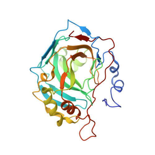
4N16

Structure of cholate bound to human carbonic anhydrase II


Experimental Data Snapshot

  • Method: X-RAY DIFFRACTION
  • Resolution: 1.54 Å
  • R-Value Free: 0.171 
  • R-Value Work: 0.145 
  • R-Value Observed: 0.146 

wwPDB Validation   3D Report Full Report


Ligand Structure Quality Assessment 


This is version 1.3 of the entry. See complete history


Literature

Structural elucidation of the hormonal inhibition mechanism of the bile acid cholate on human carbonic anhydrase II.

Boone, C.D.Tu, C.McKenna, R.

(2014) Acta Crystallogr D Biol Crystallogr 70: 1758-1763

  • DOI: https://doi.org/10.1107/S1399004714007457
  • Primary Citation of Related Structures:  
    4N16

  • PubMed Abstract: 

    The carbonic anhydrases (CAs) are a family of mostly zinc metalloenzymes that catalyze the reversible hydration/dehydration of CO2 into bicarbonate and a proton. Human isoform CA II (HCA II) is abundant in the surface epithelial cells of the gastric mucosa, where it serves an important role in cytoprotection through bicarbonate secretion. Physiological inhibition of HCA II via the bile acids contributes to mucosal injury in ulcerogenic conditions. This study details the weak biophysical interactions associated with the binding of a primary bile acid, cholate, to HCA II. The X-ray crystallographic structure determined to 1.54 Å resolution revealed that cholate does not make any direct hydrogen-bond interactions with HCA II, but instead reconfigures the well ordered water network within the active site to promote indirect binding to the enzyme. Structural knowledge of the binding interactions of this nonsulfur-containing inhibitor with HCA II could provide the template design for high-affinity, isoform-specific therapeutic agents for a variety of diseases/pathological states, including cancer, glaucoma, epilepsy and osteoporosis.


  • Organizational Affiliation

    Department of Biochemistry and Molecular Biology, University of Florida, PO Box 100267, Gainesville, FL 32610, USA.


Macromolecules
Find similar proteins by:  (by identity cutoff)  |  3D Structure
Entity ID: 1
MoleculeChains Sequence LengthOrganismDetailsImage
Carbonic anhydrase 2260Homo sapiensMutation(s): 0 
Gene Names: CA2
EC: 4.2.1.1 (PDB Primary Data), 4.2.1.69 (UniProt)
UniProt & NIH Common Fund Data Resources
Find proteins for P00918 (Homo sapiens)
Explore P00918 
Go to UniProtKB:  P00918
PHAROS:  P00918
GTEx:  ENSG00000104267 
Entity Groups  
Sequence Clusters30% Identity50% Identity70% Identity90% Identity95% Identity100% Identity
UniProt GroupP00918
Sequence Annotations
Expand
  • Reference Sequence
Small Molecules
Ligands 3 Unique
IDChains Name / Formula / InChI Key2D Diagram3D Interactions
CHD
Query on CHD

Download Ideal Coordinates CCD File 
B [auth A]CHOLIC ACID
C24 H40 O5
BHQCQFFYRZLCQQ-OELDTZBJSA-N
GOL
Query on GOL

Download Ideal Coordinates CCD File 
C [auth A],
D [auth A],
E [auth A],
F [auth A],
G [auth A]
GLYCEROL
C3 H8 O3
PEDCQBHIVMGVHV-UHFFFAOYSA-N
ZN
Query on ZN

Download Ideal Coordinates CCD File 
H [auth A]ZINC ION
Zn
PTFCDOFLOPIGGS-UHFFFAOYSA-N
Experimental Data & Validation

Experimental Data

  • Method: X-RAY DIFFRACTION
  • Resolution: 1.54 Å
  • R-Value Free: 0.171 
  • R-Value Work: 0.145 
  • R-Value Observed: 0.146 
  • Space Group: P 1 21 1
Unit Cell:
Length ( Å )Angle ( ˚ )
a = 42.358α = 90
b = 41.595β = 104.53
c = 71.931γ = 90
Software Package:
Software NamePurpose
DENZOdata reduction
SCALEPACKdata scaling
PHENIXrefinement
PDB_EXTRACTdata extraction
CrystalCleardata collection
HKL-2000data reduction
HKL-3000data scaling
PHENIXphasing

Structure Validation

View Full Validation Report


Ligand Structure Quality Assessment 


Entry History 

Deposition Data

Revision History  (Full details and data files)

  • Version 1.0: 2014-06-18
    Type: Initial release
  • Version 1.1: 2014-09-24
    Changes: Database references
  • Version 1.2: 2017-11-15
    Changes: Refinement description
  • Version 1.3: 2024-02-28
    Changes: Data collection, Database references, Derived calculations