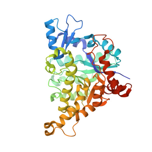
RCSB PDB - 4LAL: Crystal structure of Cordyceps militaris IDCase D323A mutant in complex with 5-carboxyl-uracil

 4LAL

Crystal structure of Cordyceps militaris IDCase D323A mutant in complex with 5-carboxyl-uracil


Experimental Data Snapshot

  • Method: X-RAY DIFFRACTION
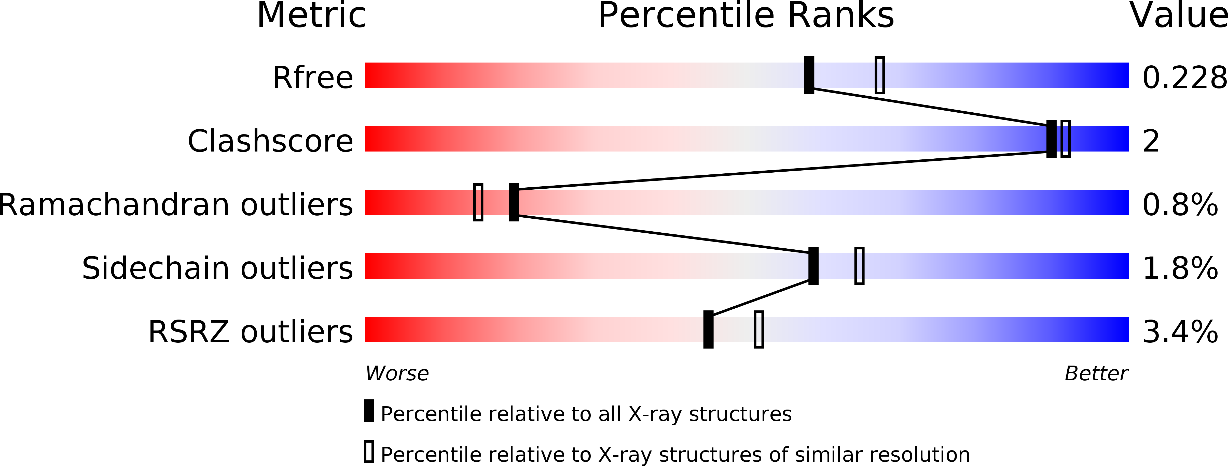
  • Resolution: 2.10 Å
  • R-Value Free: 0.220 
  • R-Value Work: 0.175 
  • R-Value Observed: 0.177 

Starting Model: experimental
View more details

wwPDB Validation   3D Report Full Report


Ligand Structure Quality Assessment 

Created with Raphaël 2.3.0Worse 01 BetterLigand structure goodness of fit to experimental dataBest fitted 5CUClick on this verticalbar to view details

This is version 1.3 of the entry. See complete history


Literature

Crystal structures of isoorotate decarboxylases reveal a novel catalytic mechanism of 5-carboxyl-uracil decarboxylation and shed light on the search for DNA decarboxylase.

Xu, S.Li, W.Zhu, J.Wang, R.Li, Z.Xu, G.L.Ding, J.

(2013) Cell Res 23: 1296-1309

  • DOI: https://doi.org/10.1038/cr.2013.107
  • Primary Citation of Related Structures:  
    4HJW, 4HK5, 4HK6, 4HK7, 4LAK, 4LAL, 4LAM, 4LAN, 4LAO

  • PubMed Abstract: 

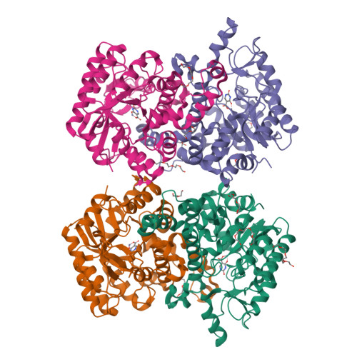
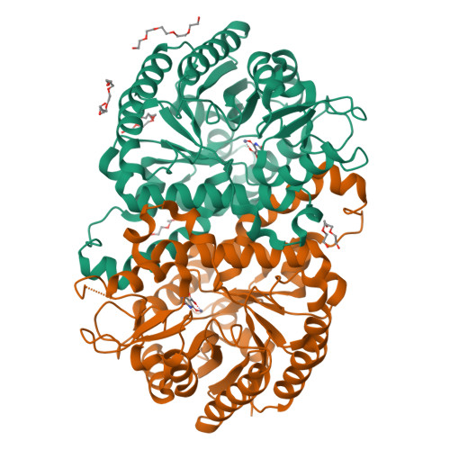
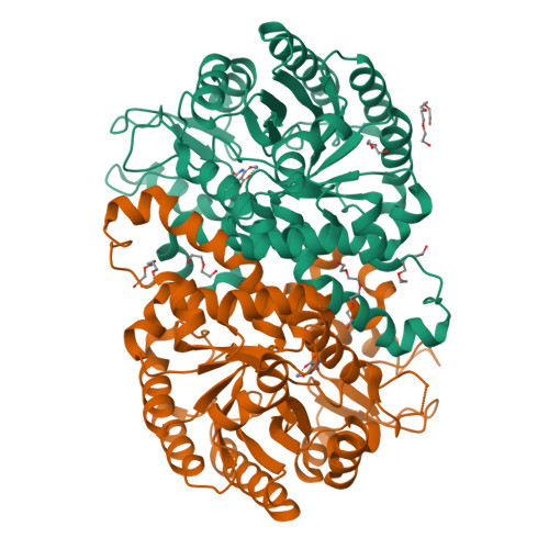
    DNA methylation and demethylation regulate many crucial biological processes in mammals and are linked to many diseases. Active DNA demethylation is believed to be catalyzed by TET proteins and a putative DNA decarboxylase that may share some similarities in sequence, structure and catalytic mechanism with isoorotate decarboxylase (IDCase) that catalyzes decarboxylation of 5caU to U in fungi. We report here the structures of wild-type and mutant IDCases from Cordyceps militaris and Metarhizium anisopliae in apo form or in complexes with 5caU, U, and an inhibitor 5-nitro-uracil. IDCases adopt a typical (β/α)8 barrel fold of the amidohydrolase superfamily and function as dimers. A Zn(2+) is bound at the active site and coordinated by four strictly conserved residues, one Asp and three His. The substrate is recognized by several strictly conserved residues. The functional roles of the key residues at the active site are validated by mutagenesis and biochemical studies. Based on the structural and biochemical data, we present for the first time a novel catalytic mechanism of decarboxylation for IDCases, which might also apply to other members of the amidohydrolase superfamily. In addition, our biochemical data show that IDCases can catalyze decarboxylation of 5caC to C albeit with weak activity, which is the first in vitro evidence for direct decarboxylation of 5caC to C by an enzyme. These findings are valuable in the identification of potential DNA decarboxylase in mammals.


  • Organizational Affiliation

    1] State Key Laboratory of Molecular Biology, Institute of Biochemistry and Cell Biology, Shanghai Institutes for Biological Sciences, Chinese Academy of Sciences, 320 Yue-Yang Road, Shanghai 200031, China [2] Graduate School of Chinese Academy of Sciences, 320 Yue-Yang Road, Shanghai 200031, China.


Macromolecules
Find similar proteins by:  (by identity cutoff)  |  3D Structure
Entity ID: 1
MoleculeChains Sequence LengthOrganismDetailsImage
Uracil-5-carboxylate decarboxylase
A, B, C, D
376Cordyceps militaris CM01Mutation(s): 1 
Gene Names: CCM_01452
UniProt
Find proteins for G3J531 (Cordyceps militaris (strain CM01))
Explore G3J531 
Go to UniProtKB:  G3J531
Entity Groups  
Sequence Clusters30% Identity50% Identity70% Identity90% Identity95% Identity100% Identity
UniProt GroupG3J531
Sequence Annotations
Expand
  • Reference Sequence
Small Molecules
Ligands 3 Unique
IDChains Name / Formula / InChI Key2D Diagram3D Interactions
P6G
Query on P6G

Download Ideal Coordinates CCD File 
G [auth A]
H [auth A]
I [auth A]
J [auth A]
M [auth B]
HEXAETHYLENE GLYCOL
C12 H26 O7
IIRDTKBZINWQAW-UHFFFAOYSA-N
5CU
Query on 5CU

Download Ideal Coordinates CCD File 
F [auth A],
L [auth B],
O [auth C],
U [auth D]
2,4-dioxo-1,2,3,4-tetrahydropyrimidine-5-carboxylic acid
C5 H4 N2 O4
ZXYAAVBXHKCJJB-UHFFFAOYSA-N
ZN
Query on ZN

Download Ideal Coordinates CCD File 
E [auth A],
K [auth B],
N [auth C],
T [auth D]
ZINC ION
Zn
PTFCDOFLOPIGGS-UHFFFAOYSA-N
Experimental Data & Validation

Experimental Data

  • Method: X-RAY DIFFRACTION
  • Resolution: 2.10 Å
  • R-Value Free: 0.220 
  • R-Value Work: 0.175 
  • R-Value Observed: 0.177 
  • Space Group: C 1 2 1
Unit Cell:
Length ( Å )Angle ( ˚ )
a = 192.266α = 90
b = 54.301β = 121.04
c = 143.776γ = 90
Software Package:
Software NamePurpose
DENZOdata reduction
SCALEPACKdata scaling
REFMACrefinement
PDB_EXTRACTdata extraction
HKL-2000data collection
HKL-2000data reduction
PHENIXphasing

Structure Validation

View Full Validation Report


Ligand Structure Quality Assessment 

Created with Raphaël 2.3.0Worse 01 BetterLigand structure goodness of fit to experimental dataBest fitted 5CUClick on this verticalbar to view details

Entry History 

Deposition Data

Revision History  (Full details and data files)

  • Version 1.0: 2013-10-02
    Type: Initial release
  • Version 1.1: 2014-02-12
    Changes: Database references
  • Version 1.2: 2017-11-15
    Changes: Refinement description
  • Version 1.3: 2023-11-08
    Changes: Data collection, Database references, Derived calculations, Refinement description