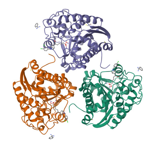
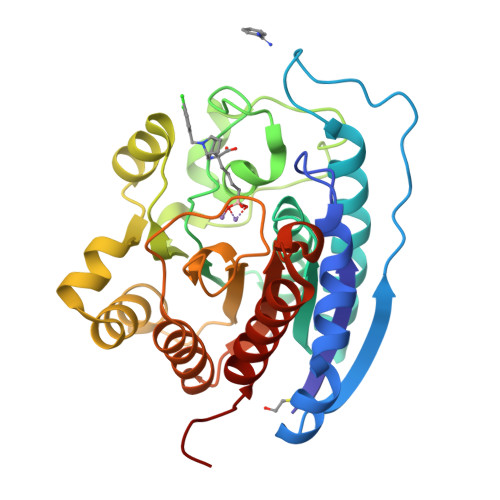
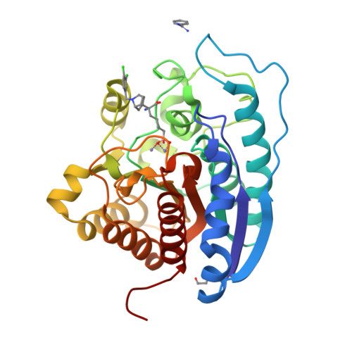
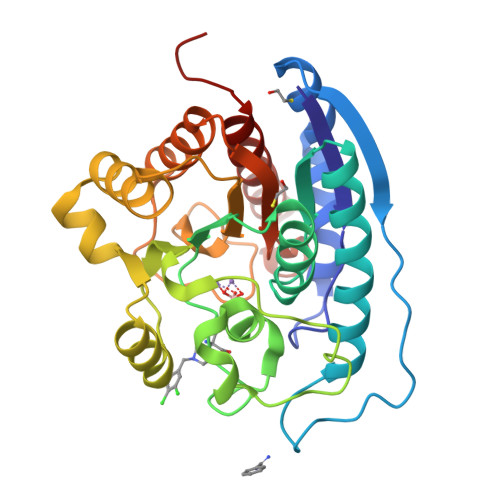
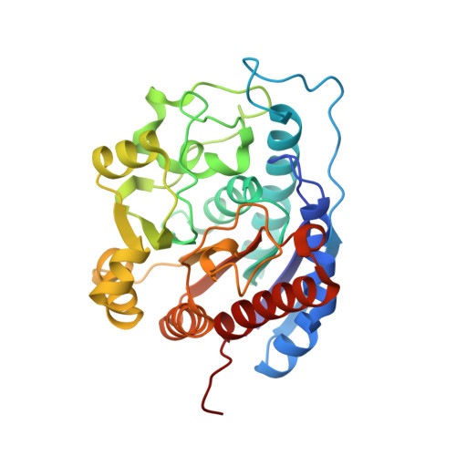
RCSB PDB - 4IXU: Crystal structure of human Arginase-2 complexed with inhibitor 11d: {(5R)-5-amino-5-carboxy-5-[(3-endo)-8-(3,4-dichlorobenzyl)-8-azabicyclo[3.2.1]oct-3-yl]pentyl}(trihydroxy)borate(1-)

 4IXU

Crystal structure of human Arginase-2 complexed with inhibitor 11d: {(5R)-5-amino-5-carboxy-5-[(3-endo)-8-(3,4-dichlorobenzyl)-8-azabicyclo[3.2.1]oct-3-yl]pentyl}(trihydroxy)borate(1-)


Experimental Data Snapshot

  • Method: X-RAY DIFFRACTION
  • Resolution: 1.90 Å
  • R-Value Free: 0.193 
  • R-Value Work: 0.167 
  • R-Value Observed: 0.169 

Starting Model: experimental
View more details

wwPDB Validation   3D Report Full Report


Ligand Structure Quality Assessment 

Created with Raphaël 2.3.0Worse 01 BetterLigand structure goodness of fit to experimental dataBest fitted 38IClick on this verticalbar to view details

This is version 1.1 of the entry. See complete history


Literature

Synthesis of quaternary alpha-amino acid-based arginase inhibitors via the Ugi reaction.

Golebiowski, A.Whitehouse, D.Beckett, R.P.Van Zandt, M.Ji, M.K.Ryder, T.R.Jagdmann, E.Andreoli, M.Lee, Y.Sheeler, R.Conway, B.Olczak, J.Mazur, M.Czestkowski, W.Piotrowska, W.Cousido-Siah, A.Ruiz, F.X.Mitschler, A.Podjarny, A.Schroeter, H.

(2013) Bioorg Med Chem Lett 23: 4837-4841

  • DOI: https://doi.org/10.1016/j.bmcl.2013.06.092
  • Primary Citation of Related Structures:  
    4IXU, 4IXV

  • PubMed Abstract: 

    The Ugi reaction has been successfully applied to the synthesis of novel arginase inhibitors. In an effort to decrease conformational flexibility of the previously reported series of 2-amino-6-boronohexanoic acid (ABH) analogs 1, we designed and synthesized a series of compounds, 2, in which a piperidine ring is linked directly to a quaternary amino acid center. Further improvement of in vitro activity was achieved by adding two carbon bridge in the piperidine ring, that is, tropane analogs 11. These improvements in activity are rationalized by X-ray crystallography analysis, which show that the tropane ring nitrogen atom moves into direct contact with Asp202 (arginase II numbering). The synthetic routes described here enabled the design of novel arginase inhibitors with improved potency and markedly different physico-chemical properties compared to ABH. Compound 11c represents the most in vitro active arginase inhibitor reported to date.


  • Organizational Affiliation

    Institutes for Pharmaceutical Discovery, 23 Business Park Drive, Branford, CT, United States. golebiowski.a@gmail.com


Macromolecules
Find similar proteins by:  (by identity cutoff)  |  3D Structure
Entity ID: 1
MoleculeChains Sequence LengthOrganismDetailsImage
Arginase-2, mitochondrial
A, B, C
306Homo sapiensMutation(s): 0 
Gene Names: ARG2
EC: 3.5.3.1
UniProt & NIH Common Fund Data Resources
Find proteins for P78540 (Homo sapiens)
Explore P78540 
Go to UniProtKB:  P78540
PHAROS:  P78540
GTEx:  ENSG00000081181 
Entity Groups  
Sequence Clusters30% Identity50% Identity70% Identity90% Identity95% Identity100% Identity
UniProt GroupP78540
Sequence Annotations
Expand
  • Reference Sequence
Small Molecules
Ligands 4 Unique
IDChains Name / Formula / InChI Key2D Diagram3D Interactions
38I
Query on 38I

Download Ideal Coordinates CCD File 
I [auth A],
O [auth B],
U [auth C]
{(5R)-5-amino-5-carboxy-5-[(3-endo)-8-(3,4-dichlorobenzyl)-8-azabicyclo[3.2.1]oct-3-yl]pentyl}(trihydroxy)borate(1-)
C20 H30 B Cl2 N2 O5
KCKQBVVBSIZAMB-UIVXKWKOSA-N
BEN
Query on BEN

Download Ideal Coordinates CCD File 
F [auth A],
L [auth B],
R [auth C]
BENZAMIDINE
C7 H8 N2
PXXJHWLDUBFPOL-UHFFFAOYSA-N
BME
Query on BME

Download Ideal Coordinates CCD File 
G [auth A]
H [auth A]
M [auth B]
N [auth B]
S [auth C]
BETA-MERCAPTOETHANOL
C2 H6 O S
DGVVWUTYPXICAM-UHFFFAOYSA-N
MN
Query on MN

Download Ideal Coordinates CCD File 
D [auth A]
E [auth A]
J [auth B]
K [auth B]
P [auth C]
MANGANESE (II) ION
Mn
WAEMQWOKJMHJLA-UHFFFAOYSA-N
Binding Affinity Annotations 
IDSourceBinding Affinity
38I PDBBind:  4IXU IC50: 23 (nM) from 1 assay(s)
Experimental Data & Validation

Experimental Data

  • Method: X-RAY DIFFRACTION
  • Resolution: 1.90 Å
  • R-Value Free: 0.193 
  • R-Value Work: 0.167 
  • R-Value Observed: 0.169 
  • Space Group: P 42 21 2
Unit Cell:
Length ( Å )Angle ( ˚ )
a = 127.4α = 90
b = 127.4β = 90
c = 159.249γ = 90
Software Package:
Software NamePurpose
PHENIXrefinement
PDB_EXTRACTdata extraction
HKL-2000data collection
HKL-2000data reduction
HKL-2000data scaling
AMoREphasing

Structure Validation

View Full Validation Report


Ligand Structure Quality Assessment 

Created with Raphaël 2.3.0Worse 01 BetterLigand structure goodness of fit to experimental dataBest fitted 38IClick on this verticalbar to view details

Entry History 

Revision History  (Full details and data files)

  • Version 1.0: 2013-12-11
    Type: Initial release
  • Version 1.1: 2023-09-20
    Changes: Data collection, Database references, Derived calculations, Refinement description