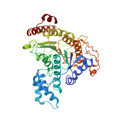
4BZ7

Crystal structure of Schistosoma mansoni HDAC8 complexed with M344


Experimental Data Snapshot

  • Method: X-RAY DIFFRACTION
  • Resolution: 1.65 Å
  • R-Value Free: 0.172 
  • R-Value Work: 0.155 
  • R-Value Observed: 0.155 

Starting Model: experimental
View more details

wwPDB Validation   3D Report Full Report


Ligand Structure Quality Assessment 


This is version 1.2 of the entry. See complete history


Literature

Structural Basis for the Inhibition of Histone Deacetylase 8 (Hdac8), a Key Epigenetic Player in the Blood Fluke Schistosoma Mansoni.

Marek, M.Kannan, S.Hauser, A.Moraes Mourao, M.Caby, S.Cura, V.Stolfa, D.A.Schmidtkunz, K.Lancelot, J.Andrade, L.Renaud, J.Oliveira, G.Sippl, W.Jung, M.Cavarelli, J.Pierce, R.J.Romier, C.

(2013) PLoS Pathog 9: 03645

  • DOI: https://doi.org/10.1371/journal.ppat.1003645
  • Primary Citation of Related Structures:  
    4BZ5, 4BZ6, 4BZ7, 4BZ8, 4BZ9

  • PubMed Abstract: 

    The treatment of schistosomiasis, a disease caused by blood flukes parasites of the Schistosoma genus, depends on the intensive use of a single drug, praziquantel, which increases the likelihood of the development of drug-resistant parasite strains and renders the search for new drugs a strategic priority. Currently, inhibitors of human epigenetic enzymes are actively investigated as novel anti-cancer drugs and have the potential to be used as new anti-parasitic agents. Here, we report that Schistosoma mansoni histone deacetylase 8 (smHDAC8), the most expressed class I HDAC isotype in this organism, is a functional acetyl-L-lysine deacetylase that plays an important role in parasite infectivity. The crystal structure of smHDAC8 shows that this enzyme adopts a canonical α/β HDAC fold, with specific solvent exposed loops corresponding to insertions in the schistosome HDAC8 sequence. Importantly, structures of smHDAC8 in complex with generic HDAC inhibitors revealed specific structural changes in the smHDAC8 active site that cannot be accommodated by human HDACs. Using a structure-based approach, we identified several small-molecule inhibitors that build on these specificities. These molecules exhibit an inhibitory effect on smHDAC8 but show reduced affinity for human HDACs. Crucially, we show that a newly identified smHDAC8 inhibitor has the capacity to induce apoptosis and mortality in schistosomes. Taken together, our biological and structural findings define the framework for the rational design of small-molecule inhibitors specifically interfering with schistosome epigenetic mechanisms, and further support an anti-parasitic epigenome targeting strategy to treat neglected diseases caused by eukaryotic pathogens.


  • Organizational Affiliation

    Département de Biologie Structurale Intégrative, Institut de Génétique et Biologie Moléculaire et Cellulaire (IGBMC), Université de Strasbourg (UDS), CNRS, INSERM, Illkirch, France.


Macromolecules
Find similar proteins by:  (by identity cutoff)  |  3D Structure
Entity ID: 1
MoleculeChains Sequence LengthOrganismDetailsImage
HISTONE DEACETYLASE 8
A, B, C, D
446Schistosoma mansoniMutation(s): 0 
EC: 3.5.1.98
UniProt
Find proteins for A5H660 (Schistosoma mansoni)
Explore A5H660 
Go to UniProtKB:  A5H660
Entity Groups  
Sequence Clusters30% Identity50% Identity70% Identity90% Identity95% Identity100% Identity
UniProt GroupA5H660
Sequence Annotations
Expand
  • Reference Sequence
Small Molecules
Ligands 4 Unique
IDChains Name / Formula / InChI Key2D Diagram3D Interactions
B3N
Query on B3N

Download Ideal Coordinates CCD File 
GA [auth D]
H [auth A]
HA [auth D]
I [auth A]
P [auth B]
GA [auth D],
H [auth A],
HA [auth D],
I [auth A],
P [auth B],
Q [auth B],
X [auth C],
Y [auth C]
4-(dimethylamino)-N-[7-(hydroxyamino)-7-oxoheptyl]benzamide
C16 H25 N3 O3
MXWDSZWTBOCWBK-UHFFFAOYSA-N
GOL
Query on GOL

Download Ideal Coordinates CCD File 
AA [auth C]
BA [auth C]
CA [auth C]
IA [auth D]
J [auth A]
AA [auth C],
BA [auth C],
CA [auth C],
IA [auth D],
J [auth A],
JA [auth D],
K [auth A],
KA [auth D],
L [auth A],
LA [auth D],
R [auth B],
S [auth B],
T [auth B],
Z [auth C]
GLYCEROL
C3 H8 O3
PEDCQBHIVMGVHV-UHFFFAOYSA-N
ZN
Query on ZN

Download Ideal Coordinates CCD File 
DA [auth D],
E [auth A],
M [auth B],
U [auth C]
ZINC ION
Zn
PTFCDOFLOPIGGS-UHFFFAOYSA-N
K
Query on K

Download Ideal Coordinates CCD File 
EA [auth D]
F [auth A]
FA [auth D]
G [auth A]
N [auth B]
EA [auth D],
F [auth A],
FA [auth D],
G [auth A],
N [auth B],
O [auth B],
V [auth C],
W [auth C]
POTASSIUM ION
K
NPYPAHLBTDXSSS-UHFFFAOYSA-N
Binding Affinity Annotations 
IDSourceBinding Affinity
B3N BindingDB:  4BZ7 IC50: 3162 (nM) from 1 assay(s)
Experimental Data & Validation

Experimental Data

  • Method: X-RAY DIFFRACTION
  • Resolution: 1.65 Å
  • R-Value Free: 0.172 
  • R-Value Work: 0.155 
  • R-Value Observed: 0.155 
  • Space Group: P 1
Unit Cell:
Length ( Å )Angle ( ˚ )
a = 70.557α = 78.05
b = 70.579β = 75.41
c = 98.513γ = 85.49
Software Package:
Software NamePurpose
BUSTERrefinement
DENZOdata reduction
SCALEPACKdata scaling
MOLREPphasing

Structure Validation

View Full Validation Report


Ligand Structure Quality Assessment 


Entry History 

Deposition Data

Revision History  (Full details and data files)

  • Version 1.0: 2013-08-14
    Type: Initial release
  • Version 1.1: 2013-10-16
    Changes: Database references
  • Version 1.2: 2023-12-20
    Changes: Data collection, Database references, Derived calculations, Other, Refinement description