3ZHP

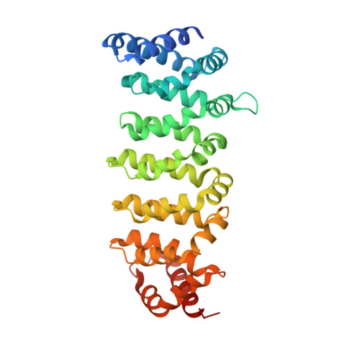
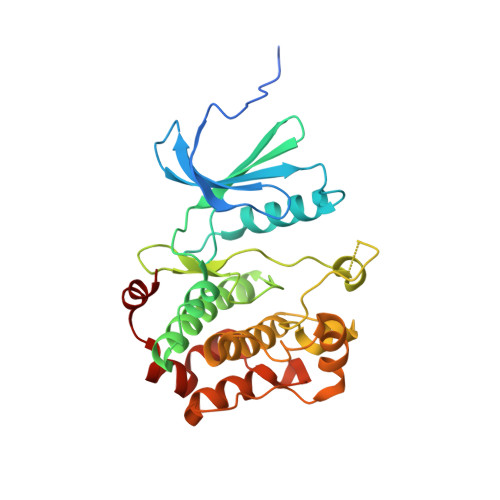
Human MST3 (STK24) in complex with MO25beta


Experimental Data Snapshot

  • Method: X-RAY DIFFRACTION
  • Resolution: 2.90 Å
  • R-Value Free: 0.261 
  • R-Value Work: 0.227 
  • R-Value Observed: 0.228 

Starting Models: experimental
View more details

wwPDB Validation   3D Report Full Report


This is version 1.4 of the entry. See complete history


Literature

Structural Insights Into the Activation of Mst3 by Mo25.

Mehellou, Y.Alessi, D.R.Macartney, T.J.Szklarz, M.Knapp, S.Elkins, J.M.

(2013) Biochem Biophys Res Commun 431: 604

  • DOI: https://doi.org/10.1016/j.bbrc.2012.12.113
  • Primary Citation of Related Structures:  
    3ZHP

  • PubMed Abstract: 

    The MO25 scaffolding protein operates as critical regulator of a number of STE20 family protein kinases (e.g. MST and SPAK isoforms) as well as pseudokinases (e.g. STRAD isoforms that play a critical role in activating the LKB1 tumour suppressor). To better understand how MO25 interacts and stimulates the activity of STE20 protein kinases, we determined the crystal structure of MST3 catalytic domain (residues 19-289) in complex with full length MO25β. The structure reveals an intricate web of interactions between MST3 and MO25β that function to stabilise the kinase domain in a closed, active, conformation even in the absence of ATP or an ATP-mimetic inhibitor. The binding mode of MO25β is reminiscent of the mechanism by which MO25α interacts with the pseudokinase STRADα. In particular we identified interface residues Tyr223 of MO25β and Glu58 and Ile71 of MST3 that when mutated prevent activation of MST3 by MO25β. These data provide molecular understanding of the mechanism by which MO25 isoforms regulates the activity of STE20 family protein kinases.


  • Organizational Affiliation

    MRC Protein Phosphorylation Unit, College of Life Sciences, University of Dundee, Dow Street, Dundee, DD1 5EH Scotland, UK. [email protected]


Macromolecules
Find similar proteins by:  (by identity cutoff)  |  3D Structure
Entity ID: 1
MoleculeChains Sequence LengthOrganismDetailsImage
CALCIUM-BINDING PROTEIN 39-LIKE
A, B
340Homo sapiensMutation(s): 0 
UniProt & NIH Common Fund Data Resources
Find proteins for Q9H9S4 (Homo sapiens)
Explore Q9H9S4 
Go to UniProtKB:  Q9H9S4
PHAROS:  Q9H9S4
GTEx:  ENSG00000102547 
Entity Groups  
Sequence Clusters30% Identity50% Identity70% Identity90% Identity95% Identity100% Identity
UniProt GroupQ9H9S4
Sequence Annotations
Expand
  • Reference Sequence
Find similar proteins by:  (by identity cutoff)  |  3D Structure
Entity ID: 2
MoleculeChains Sequence LengthOrganismDetailsImage
SERINE/THREONINE-PROTEIN KINASE 24
C, D
294Homo sapiensMutation(s): 0 
EC: 2.7.11.1
UniProt & NIH Common Fund Data Resources
Find proteins for Q9Y6E0 (Homo sapiens)
Explore Q9Y6E0 
Go to UniProtKB:  Q9Y6E0
PHAROS:  Q9Y6E0
GTEx:  ENSG00000102572 
Entity Groups  
Sequence Clusters30% Identity50% Identity70% Identity90% Identity95% Identity100% Identity
UniProt GroupQ9Y6E0
Sequence Annotations
Expand
  • Reference Sequence
Experimental Data & Validation

Experimental Data

  • Method: X-RAY DIFFRACTION
  • Resolution: 2.90 Å
  • R-Value Free: 0.261 
  • R-Value Work: 0.227 
  • R-Value Observed: 0.228 
  • Space Group: P 1 21 1
Unit Cell:
Length ( Å )Angle ( ˚ )
a = 64.8α = 90
b = 120.34β = 99.69
c = 98.91γ = 90
Software Package:
Software NamePurpose
BUSTERrefinement
MOSFLMdata reduction
SCALAdata scaling
PHASERphasing

Structure Validation

View Full Validation Report


Entry History 

Deposition Data

Revision History  (Full details and data files)

  • Version 1.0: 2013-01-16
    Type: Initial release
  • Version 1.1: 2013-04-17
    Changes: Database references
  • Version 1.2: 2014-07-16
    Changes: Structure summary
  • Version 1.3: 2018-01-24
    Changes: Structure summary
  • Version 1.4: 2023-12-20
    Changes: Data collection, Database references, Derived calculations, Other, Refinement description