3SHZ | pdb_00003shz

Crystal structure of the PDE5A1 catalytic domain in complex with novel inhibitors


Experimental Data Snapshot

  • Method: X-RAY DIFFRACTION
  • Resolution: 2.45 Å
  • R-Value Free: 
    0.245 (Depositor), 0.240 (DCC) 
  • R-Value Work: 
    0.201 (Depositor), 0.200 (DCC) 
  • R-Value Observed: 
    0.203 (Depositor) 

wwPDB Validation   3D Report Full Report


Ligand Structure Quality Assessment 

Created with Raphaël 2.3.0Worse 01 BetterLigand structure goodness of fit to experimental dataBest fitted 5COClick on this verticalbar to view details

This is version 1.2 of the entry. See complete history


Literature

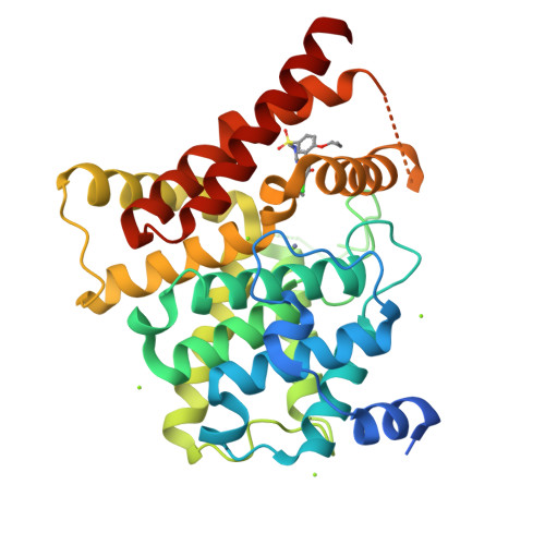
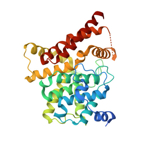
Utilization of Halogen Bond in Lead Optimization: A Case Study of Rational Design of Potent Phosphodiesterase Type 5 (PDE5) Inhibitors.

Xu, Z.Liu, Z.Chen, T.Chen, T.T.Wang, Z.Tian, G.Shi, J.Wang, X.Lu, Y.Yan, X.Wang, G.Jiang, H.Chen, K.Wang, S.Xu, Y.Shen, J.Zhu, W.

(2011) J Med Chem 54: 5607-5611

  • DOI: https://doi.org/10.1021/jm200644r
  • Primary Citation of Related Structures:  
    3SHY, 3SHZ, 3SIE

  • PubMed Abstract: 

    For proof-of-concept of halogen bonding in drug design, a series of halogenated compounds were designed based on a lead structure as new inhibitors of phosphodiesterase type 5. Bioassay results revealed a good correlation between the measured bioactivity and the calculated halogen bond energy. Our X-ray crystal structures verified the existence of the predicted halogen bonds, demonstrating that the halogen bond is an applicable tool in drug design and should be routinely considered in lead optimization.


  • Organizational Affiliation

    State Key Laboratory of Drug Research, Shanghai Institute of Materia Medica, Chinese Academy of Sciences, Shanghai, China.


Macromolecules
Find similar proteins by:  (by identity cutoff)  |  3D Structure
Entity ID: 1
MoleculeChains Sequence LengthOrganismDetailsImage
cGMP-specific 3',5'-cyclic phosphodiesterase347Homo sapiensMutation(s): 0 
Gene Names: PDE5PDE5A
EC: 3.1.4.35
UniProt & NIH Common Fund Data Resources
Find proteins for O76074 (Homo sapiens)
Explore O76074 
Go to UniProtKB:  O76074
PHAROS:  O76074
GTEx:  ENSG00000138735 
Entity Groups  
Sequence Clusters30% Identity50% Identity70% Identity90% Identity95% Identity100% Identity
UniProt GroupO76074
Sequence Annotations
Expand
  • Reference Sequence
Small Molecules
Ligands 3 Unique
IDChains Name / Formula / InChI Key2D Diagram3D Interactions
5CO
Query on 5CO

Download Ideal Coordinates CCD File 
B [auth A]5-chloro-6-ethyl-2-{5-[(4-methylpiperazin-1-yl)sulfonyl]-2-propoxyphenyl}pyrimidin-4(3H)-one
C20 H27 Cl N4 O4 S
HVJOKVUJWQTZFF-UHFFFAOYSA-N
ZN
Query on ZN

Download Ideal Coordinates CCD File 
C [auth A]ZINC ION
Zn
PTFCDOFLOPIGGS-UHFFFAOYSA-N
MG
Query on MG

Download Ideal Coordinates CCD File 
D [auth A],
E [auth A],
F [auth A],
G [auth A],
H [auth A]
MAGNESIUM ION
Mg
JLVVSXFLKOJNIY-UHFFFAOYSA-N
Binding Affinity Annotations 
IDSourceBinding Affinity
5CO BindingDB:  3SHZ Kd: 770 (nM) from 1 assay(s)
Experimental Data & Validation

Experimental Data

  • Method: X-RAY DIFFRACTION
  • Resolution: 2.45 Å
  • R-Value Free:  0.245 (Depositor), 0.240 (DCC) 
  • R-Value Work:  0.201 (Depositor), 0.200 (DCC) 
  • R-Value Observed: 0.203 (Depositor) 
Space Group: P 31 2 1
Unit Cell:
Length ( Å )Angle ( ˚ )
a = 74.687α = 90
b = 74.687β = 90
c = 131.728γ = 120
Software Package:
Software NamePurpose
DENZOdata reduction
SCALEPACKdata scaling
PHENIXrefinement
PDB_EXTRACTdata extraction
ADSCdata collection
HKL-2000data reduction
PHASERphasing

Structure Validation

View Full Validation Report


Ligand Structure Quality Assessment 

Created with Raphaël 2.3.0Worse 01 BetterLigand structure goodness of fit to experimental dataBest fitted 5COClick on this verticalbar to view details

Entry History 

Deposition Data

Revision History  (Full details and data files)

  • Version 1.0: 2011-08-24
    Type: Initial release
  • Version 1.1: 2017-11-08
    Changes: Refinement description
  • Version 1.2: 2024-03-20
    Changes: Data collection, Database references, Derived calculations