3OBT | pdb_00003obt

Crystal structure of Botulinum neurotoxin serotype D ligand binding domain in complex with N-Acetylneuraminic acid


Experimental Data Snapshot

  • Method: X-RAY DIFFRACTION
  • Resolution: 2.00 Å
  • R-Value Free: 
    0.221 (Depositor), 0.221 (DCC) 
  • R-Value Work: 
    0.172 (Depositor), 0.181 (DCC) 
  • R-Value Observed: 
    0.174 (Depositor) 

wwPDB Validation   3D Report Full Report


Ligand Structure Quality Assessment 

Created with Raphaël 2.3.0Worse 01 BetterLigand structure goodness of fit to experimental dataBest fitted SLBClick on this verticalbar to view details

This is version 1.5 of the entry. See complete history


Literature

Botulinum neurotoxin serotype D attacks neurons via two carbohydrate-binding sites in a ganglioside-dependent manner.

Strotmeier, J.Lee, K.Volker, A.K.Mahrhold, S.Zong, Y.Zeiser, J.Zhou, J.Pich, A.Bigalke, H.Binz, T.Rummel, A.Jin, R.

(2010) Biochem J 431: 207-216

  • DOI: https://doi.org/10.1042/BJ20101042
  • Primary Citation of Related Structures:  
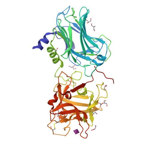
    3OBR, 3OBT

  • PubMed Abstract: 

    The extraordinarily high toxicity of botulinum neurotoxins primarily results from their specific binding and uptake into neurons. At motor neurons, the seven BoNT (botulinum neurotoxin) serotypes A-G inhibit acetylcholine release leading to flaccid paralysis. Uptake of BoNT/A, B, E, F and G requires a dual interaction with gangliosides and the synaptic vesicle proteins synaptotagmin or SV2 (synaptic vesicle glycoprotein 2), whereas little is known about the cell entry mechanisms of the serotypes C and D, which display the lowest amino acid sequence identity compared with the other five serotypes. In the present study we demonstrate that the neurotoxicity of BoNT/D depends on the presence of gangliosides by employing phrenic nerve hemidiaphragm preparations derived from mice expressing the gangliosides GM3, GM2, GM1 and GD1a, or only GM3 [a description of our use of ganglioside nomenclature is given in Svennerholm (1994) Prog. Brain Res. 101, XI-XIV]. High-resolution crystal structures of the 50 kDa cell-binding domain of BoNT/D alone and in complex with sialic acid, as well as biological analyses of single-site BoNT/D mutants identified two carbohydrate-binding sites. One site is located at a position previously identified in BoNT/A, B, E, F and G, but is lacking the conserved SXWY motif. The other site, co-ordinating one molecule of sialic acid, resembles the second ganglioside-binding pocket (the sialic-acid-binding site) of TeNT (tetanus neurotoxin).


  • Organizational Affiliation

    Institut für Toxikologie, Medizinische Hochschule Hannover, Carl-Neuberg-Strasse 1, Hannover, Germany.


Macromolecules
Find similar proteins by:  (by identity cutoff)  |  3D Structure
Entity ID: 1
MoleculeChains Sequence LengthOrganismDetailsImage
Botulinum neurotoxin type D434Clostridium botulinumMutation(s): 0 
Gene Names: botD
EC: 3.4.24.69
UniProt
Find proteins for P19321 (Clostridium botulinum D phage)
Explore P19321 
Go to UniProtKB:  P19321
Entity Groups  
Sequence Clusters30% Identity50% Identity70% Identity90% Identity95% Identity100% Identity
UniProt GroupP19321
Sequence Annotations
Expand
  • Reference Sequence
Small Molecules
Ligands 2 Unique
IDChains Name / Formula / InChI Key2D Diagram3D Interactions
SLB
Query on SLB

Download Ideal Coordinates CCD File 
B [auth A]N-acetyl-beta-neuraminic acid
C11 H19 N O9
SQVRNKJHWKZAKO-PFQGKNLYSA-N
GOL
Query on GOL

Download Ideal Coordinates CCD File 
C [auth A]
D [auth A]
E [auth A]
F [auth A]
G [auth A]
GLYCEROL
C3 H8 O3
PEDCQBHIVMGVHV-UHFFFAOYSA-N
Experimental Data & Validation

Experimental Data

  • Method: X-RAY DIFFRACTION
  • Resolution: 2.00 Å
  • R-Value Free:  0.221 (Depositor), 0.221 (DCC) 
  • R-Value Work:  0.172 (Depositor), 0.181 (DCC) 
  • R-Value Observed: 0.174 (Depositor) 
Space Group: P 21 21 21
Unit Cell:
Length ( Å )Angle ( ˚ )
a = 60.232α = 90
b = 90.044β = 90
c = 93.518γ = 90
Software Package:
Software NamePurpose
SCALEPACKdata scaling
REFMACrefinement
PDB_EXTRACTdata extraction
Blu-Icedata collection
DENZOdata reduction
PHASERphasing

Structure Validation

View Full Validation Report


Ligand Structure Quality Assessment 

Created with Raphaël 2.3.0Worse 01 BetterLigand structure goodness of fit to experimental dataBest fitted SLBClick on this verticalbar to view details

Entry History 

Deposition Data

Revision History  (Full details and data files)

  • Version 1.0: 2010-09-08
    Type: Initial release
  • Version 1.1: 2011-07-13
    Changes: Version format compliance
  • Version 1.2: 2014-04-16
    Changes: Other
  • Version 1.3: 2017-11-08
    Changes: Refinement description
  • Version 1.4: 2020-07-29
    Type: Remediation
    Reason: Carbohydrate remediation
    Changes: Data collection, Database references, Derived calculations, Structure summary
  • Version 1.5: 2024-10-30
    Changes: Data collection, Database references, Structure summary