3MSH | pdb_00003msh

Crystal structure of Hepatitis B X-Interacting Protein at high resolution


Experimental Data Snapshot

  • Method: X-RAY DIFFRACTION
  • Resolution: 1.51 Å
  • R-Value Free: 
    0.212 (Depositor) 
  • R-Value Work: 
    0.180 (Depositor), 0.180 (DCC) 
  • R-Value Observed: 
    0.183 (Depositor) 

Starting Model: experimental
View more details

wwPDB Validation   3D Report Full Report


This is version 1.3 of the entry. See complete history


Literature

Structural Characterization of HBXIP: The Protein That Interacts with the Anti-Apoptotic Protein Survivin and the Oncogenic Viral Protein HBx.

Garcia-Saez, I.Lacroix, F.B.Blot, D.Gabel, F.Skoufias, D.A.

(2011) J Mol Biology 405: 331-340

  • DOI: https://doi.org/10.1016/j.jmb.2010.10.046
  • Primary Citation of Related Structures:  
    3MS6, 3MSH

  • PubMed Abstract: 

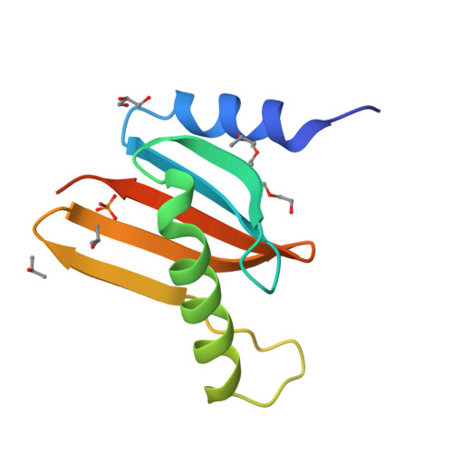
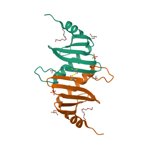
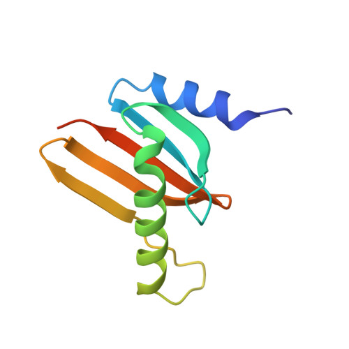
    Hepatitis B X-interacting protein (HBXIP) is a ubiquitous protein that was originally identified as a binding partner of the hepatitis B viral protein HBx. HBXIP is also thought to serve as an anti-apoptotic cofactor of survivin, promoting the suppression of pro-caspase-9 activation. Here were port the crystal structure of the shortest isoform of HBXIP (91 aa long,∼11 kDa) at 1.5 Å resolution. HBXIP crystal shows a monomer per asymmetric unit, with a profilin-like fold which is common to a super family of proteins, the Roadblock/LC7 domain family involved in protein-protein interactions. Based on this fold, we propose that HBXIP can form a dimer that can indeed be found in the crystal when symmetric molecules are generated around the asymmetric unit. This dimer shows an extended β-sheet area formed by 10 anti-parallel β-strands from both subunits. Another interesting aspect of the proposed HBXIP dimer interface is the presence of a small leucine zipper between the two α2 helices of each monomer. In solution, the scattering curve obtained by small-angle X-ray scattering for the sample used for crystallization indicates that the protein is dimeric form in solution. The fit between the experimental small angle X-ray scattering curve and the back calculated curves for two potential crystal dimers shows a significant preference for the Roadblock/LC7 fold dimer model. Moreover, the HBXIP crystal structure represents a step towards understanding the cellular role of HBXIP.


  • Organizational Affiliation

    Institut de Biologie Structurale J.-P. Ebel, UMR 5075, Grenoble Cedex 1, France. isabel.garcia@ibs.fr


Macromolecules
Find similar proteins by:  (by identity cutoff)  |  3D Structure
Entity ID: 1
MoleculeChains Sequence LengthOrganismDetailsImage
Hepatitis B virus X-interacting protein99Homo sapiensMutation(s): 0 
Gene Names: HBXIPXIP
UniProt & NIH Common Fund Data Resources
Find proteins for O43504 (Homo sapiens)
Explore O43504 
Go to UniProtKB:  O43504
PHAROS:  O43504
GTEx:  ENSG00000134248 
Entity Groups  
Sequence Clusters30% Identity50% Identity70% Identity90% Identity95% Identity100% Identity
UniProt GroupO43504
Sequence Annotations
Expand
  • Reference Sequence
Small Molecules
Ligands 4 Unique
IDChains Name / Formula / InChI Key2D Diagram3D Interactions
PG4
Query on PG4

Download Ideal Coordinates CCD File 
G [auth A]TETRAETHYLENE GLYCOL
C8 H18 O5
UWHCKJMYHZGTIT-UHFFFAOYSA-N
PO4
Query on PO4

Download Ideal Coordinates CCD File 
F [auth A]PHOSPHATE ION
O4 P
NBIIXXVUZAFLBC-UHFFFAOYSA-K
GOL
Query on GOL

Download Ideal Coordinates CCD File 
B [auth A]GLYCEROL
C3 H8 O3
PEDCQBHIVMGVHV-UHFFFAOYSA-N
IPA
Query on IPA

Download Ideal Coordinates CCD File 
C [auth A],
D [auth A],
E [auth A]
ISOPROPYL ALCOHOL
C3 H8 O
KFZMGEQAYNKOFK-UHFFFAOYSA-N
Experimental Data & Validation

Experimental Data

  • Method: X-RAY DIFFRACTION
  • Resolution: 1.51 Å
  • R-Value Free:  0.212 (Depositor) 
  • R-Value Work:  0.180 (Depositor), 0.180 (DCC) 
  • R-Value Observed: 0.183 (Depositor) 
Space Group: P 41 21 2
Unit Cell:
Length ( Å )Angle ( ˚ )
a = 49.161α = 90
b = 49.161β = 90
c = 72.195γ = 90
Software Package:
Software NamePurpose
DNAdata collection
PHASERphasing
PHENIXrefinement
XDSdata reduction
SCALAdata scaling

Structure Validation

View Full Validation Report


Entry History 

Deposition Data

Revision History  (Full details and data files)

  • Version 1.0: 2010-11-10
    Type: Initial release
  • Version 1.1: 2011-07-13
    Changes: Version format compliance
  • Version 1.2: 2023-09-06
    Changes: Data collection, Database references, Derived calculations, Refinement description
  • Version 1.3: 2024-11-20
    Changes: Structure summary