3LSS | pdb_00003lss

Trypanosoma brucei seryl-tRNA synthetase in complex with ATP


Experimental Data Snapshot

  • Method: X-RAY DIFFRACTION
  • Resolution: 1.95 Å
  • R-Value Free: 
    0.195 (Depositor), 0.200 (DCC) 
  • R-Value Work: 
    0.168 (Depositor), 0.180 (DCC) 
  • R-Value Observed: 
    0.169 (Depositor) 

Starting Model: experimental
View more details

wwPDB Validation   3D Report Full Report


Ligand Structure Quality Assessment 

Created with Raphaël 2.3.0Worse 01 BetterLigand structure goodness of fit to experimental dataBest fitted ATPClick on this verticalbar to view details

This is version 1.3 of the entry. See complete history


Literature

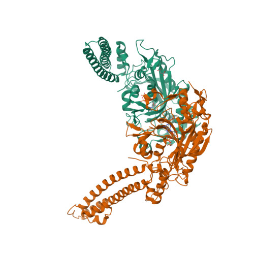
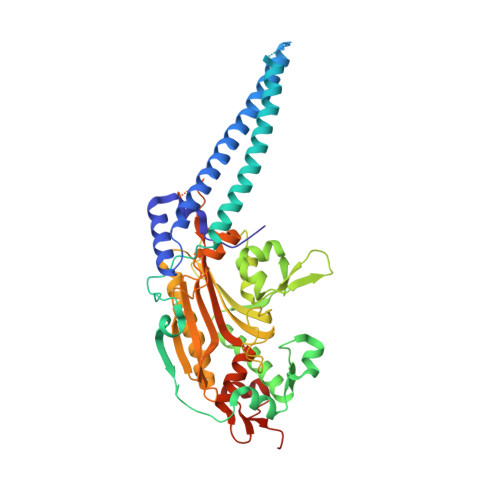
X-ray crystal structure of Seryl-tRNA synthetase from the eukaryotic parasite Trypanosoma brucei.

Larson, E.T.Zhang, L.Napuli, A.Mueller, N.Verlinde, C.L.M.J.Van Voorhis, W.C.Buckner, F.S.Fan, E.Hol, W.G.J.Merritt, E.A.

To be published.

Macromolecules
Find similar proteins by:  (by identity cutoff)  |  3D Structure
Entity ID: 1
MoleculeChains Sequence LengthOrganismDetailsImage
Seryl-tRNA synthetase
A, B
484Trypanosoma bruceiMutation(s): 0 
Gene Names: SerRSTb11.02.5020
EC: 6.1.1.11
UniProt
Find proteins for Q384V4 (Trypanosoma brucei brucei (strain 927/4 GUTat10.1))
Explore Q384V4 
Go to UniProtKB:  Q384V4
Entity Groups  
Sequence Clusters30% Identity50% Identity70% Identity90% Identity95% Identity100% Identity
UniProt GroupQ384V4
Sequence Annotations
Expand
  • Reference Sequence
Experimental Data & Validation

Experimental Data

  • Method: X-RAY DIFFRACTION
  • Resolution: 1.95 Å
  • R-Value Free:  0.195 (Depositor), 0.200 (DCC) 
  • R-Value Work:  0.168 (Depositor), 0.180 (DCC) 
  • R-Value Observed: 0.169 (Depositor) 
Space Group: H 3 2
Unit Cell:
Length ( Å )Angle ( ˚ )
a = 175.652α = 90
b = 175.652β = 90
c = 248.518γ = 120
Software Package:
Software NamePurpose
DENZOdata reduction
SCALEPACKdata scaling
REFMACrefinement
PDB_EXTRACTdata extraction
Blu-Icedata collection
HKL-2000data reduction
HKL-2000data scaling
REFMACphasing

Structure Validation

View Full Validation Report


Ligand Structure Quality Assessment 

Created with Raphaël 2.3.0Worse 01 BetterLigand structure goodness of fit to experimental dataBest fitted ATPClick on this verticalbar to view details

Entry History 

Deposition Data

Revision History  (Full details and data files)

  • Version 1.0: 2010-03-02
    Type: Initial release
  • Version 1.1: 2011-07-13
    Changes: Version format compliance
  • Version 1.2: 2017-11-01
    Changes: Refinement description
  • Version 1.3: 2023-09-06
    Changes: Data collection, Database references, Derived calculations, Refinement description