3LO8 | pdb_00003lo8

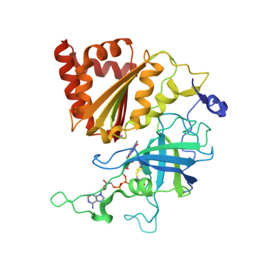
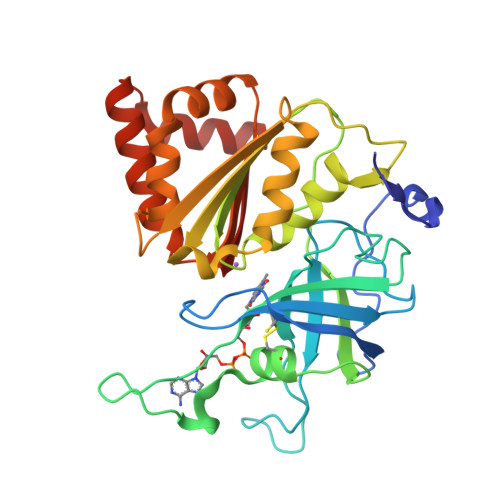
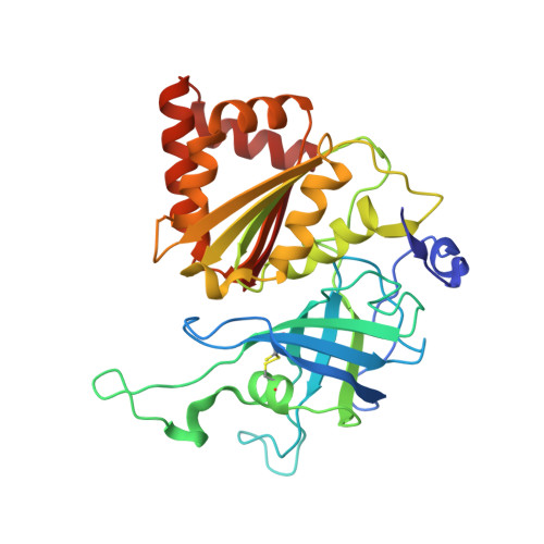
Crystal Structure of The Oxidized Form of Ferredoxin:NADP+ Reductase From Maize Root at 1.05 Angstroms


Experimental Data Snapshot

  • Method: X-RAY DIFFRACTION
  • Resolution: 1.05 Å
  • R-Value Free: 
    0.155 (Depositor), 0.160 (DCC) 
  • R-Value Work: 
    0.125 (Depositor), 0.130 (DCC) 
  • R-Value Observed: 
    0.125 (Depositor) 

Starting Model: experimental
View more details

wwPDB Validation   3D Report Full Report


Ligand Structure Quality Assessment 

Created with Raphaël 2.3.0Worse 01 BetterLigand structure goodness of fit to experimental dataBest fitted FADClick on this verticalbar to view details

This is version 1.4 of the entry. See complete history


Literature

Using a conformation-dependent stereochemical library improves crystallographic refinement of proteins.

Tronrud, D.E.Berkholz, D.S.Karplus, P.A.

(2010) Acta Crystallogr D Biol Crystallogr 66: 834-842

  • DOI: https://doi.org/10.1107/S0907444910019207
  • Primary Citation of Related Structures:  
    3LO8

  • PubMed Abstract: 

    The major macromolecular crystallographic refinement packages restrain models to ideal geometry targets defined as single values that are independent of molecular conformation. However, ultrahigh-resolution X-ray models of proteins are not consistent with this concept of ideality and have been used to develop a library of ideal main-chain bond lengths and angles that are parameterized by the phi/psi angle of the residue [Berkholz et al. (2009), Structure, 17, 1316-1325]. Here, it is first shown that the new conformation-dependent library does not suffer from poor agreement with ultrahigh-resolution structures, whereas current libraries have this problem. Using the TNT refinement package, it is then shown that protein structure refinement using this conformation-dependent library results in models that have much better agreement with library values of bond angles with little change in the R values. These tests support the value of revising refinement software to account for this new paradigm.


  • Organizational Affiliation

    Department of Biophysics and Biochemistry, Oregon State University, Corvallis, Oregon 97331, USA.


Macromolecules
Find similar proteins by:  (by identity cutoff)  |  3D Structure
Entity ID: 1
MoleculeChains Sequence LengthOrganismDetailsImage
Ferredoxin--NADP reductase311Zea maysMutation(s): 0 
EC: 1.18.1.2
UniProt
Find proteins for Q41736 (Zea mays)
Explore Q41736 
Go to UniProtKB:  Q41736
Entity Groups  
Sequence Clusters30% Identity50% Identity70% Identity90% Identity95% Identity100% Identity
UniProt GroupQ41736
Sequence Annotations
Expand
  • Reference Sequence
Experimental Data & Validation

Experimental Data

  • Method: X-RAY DIFFRACTION
  • Resolution: 1.05 Å
  • R-Value Free:  0.155 (Depositor), 0.160 (DCC) 
  • R-Value Work:  0.125 (Depositor), 0.130 (DCC) 
  • R-Value Observed: 0.125 (Depositor) 
Space Group: P 32 2 1
Unit Cell:
Length ( Å )Angle ( ˚ )
a = 59.14α = 90
b = 59.14β = 90
c = 186.74γ = 120
Software Package:
Software NamePurpose
SHELXL-97refinement
TNTrefinement
DENZOdata reduction
SCALEPACKdata scaling
TNTphasing

Structure Validation

View Full Validation Report


Ligand Structure Quality Assessment 

Created with Raphaël 2.3.0Worse 01 BetterLigand structure goodness of fit to experimental dataBest fitted FADClick on this verticalbar to view details

Entry History 

Deposition Data

Revision History  (Full details and data files)

  • Version 1.0: 2010-03-16
    Type: Initial release
  • Version 1.1: 2011-07-13
    Changes: Version format compliance
  • Version 1.2: 2019-07-17
    Changes: Data collection, Refinement description
  • Version 1.3: 2023-09-06
    Changes: Data collection, Database references, Derived calculations, Refinement description
  • Version 1.4: 2024-10-16
    Changes: Structure summary