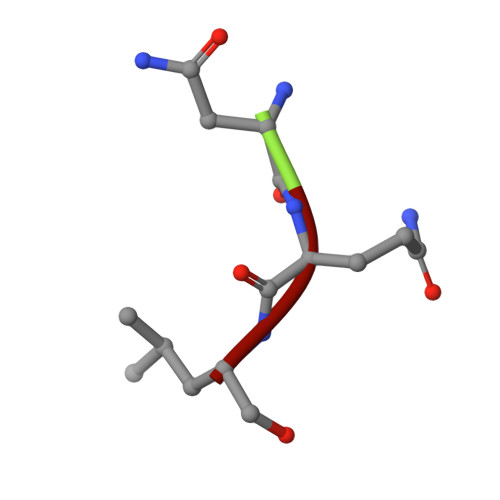
3KRD | pdb_00003krd

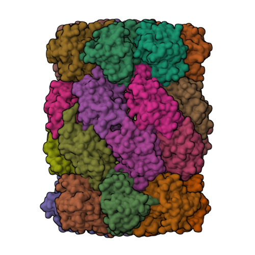
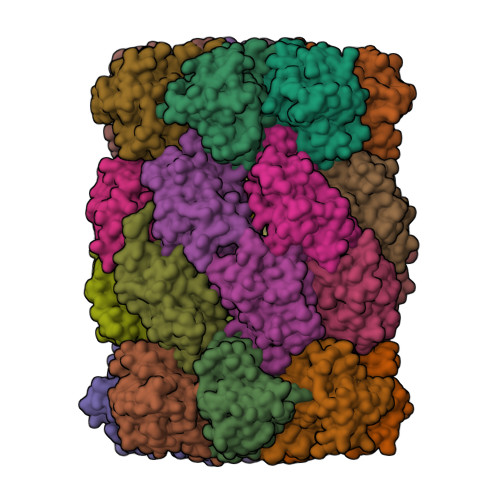
Crystal Structure of Mycobacterium Tuberculosis Proteasome in complex with Fellutamide B


Experimental Data Snapshot

  • Method: X-RAY DIFFRACTION
  • Resolution: 2.50 Å
  • R-Value Free: 
    0.229 (Depositor), 0.220 (DCC) 
  • R-Value Work: 
    0.208 (Depositor), 0.200 (DCC) 
  • R-Value Observed: 
    0.208 (Depositor) 

wwPDB Validation   3D Report Full Report


Ligand Structure Quality Assessment 

Created with Raphaël 2.3.0Worse 01 BetterLigand structure goodness of fit to experimental dataBest fitted HXDClick on this verticalbar to view details

This is version 2.1 of the entry. See complete history


Literature

Fellutamide B is a potent inhibitor of the Mycobacterium tuberculosis proteasome.

Lin, G.Li, D.Chidawanyika, T.Nathan, C.Li, H.

(2010) Arch Biochem Biophys 501: 214-220

  • DOI: https://doi.org/10.1016/j.abb.2010.06.009
  • Primary Citation of Related Structures:  
    3KRD

  • PubMed Abstract: 

    Via high-throughput screening of a natural compound library, we have identified a lipopeptide aldehyde, fellutamide B (1), as the most potent inhibitor of the Mycobacterium tuberculosis (Mtb) proteasome tested to date. Kinetic studies reveal that 1 inhibits both Mtb and human proteasomes in a time-dependent manner under steady-state condition. Remarkably, 1 inhibits the Mtb proteasome in a single-step binding mechanism with K(i)=6.8 nM, whereas it inhibits the human proteasome beta5 active site following a two-step mechanism with K(i)=11.5 nM and K(i)(*)=0.93 nM. Co-crystallization of 1 bound to the Mtb proteasome revealed a structural basis for the tight binding of 1 to the active sites of the Mtb proteasome. The hemiacetal group of 1 in the Mtb proteasome takes the (R)-configuration, whereas in the yeast proteasome it takes the (S)-configuration, indicating that the pre-chiral CHO group of 1 binds to the active site Thr1 in a different orientation. Re-examination of the structure of the yeast proteasome in complex with 1 showed significant conformational changes at the substrate-binding cleft along the active site. These structural differences are consistent with the different kinetic mechanisms of 1 against Mtb and human proteasomes.


  • Organizational Affiliation

    Department of Microbiology and Immunology, Weill Medical College of Cornell University, 1300 York Ave., New York, NY 10065, United States. gal2005@med.cornell.edu


Macromolecules
Find similar proteins by:  (by identity cutoff)  |  3D Structure
Entity ID: 1
MoleculeChains Sequence LengthOrganismDetailsImage
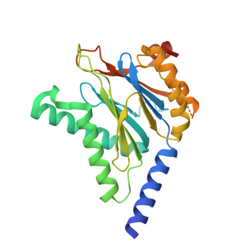
Proteasome subunit alpha248Mycobacterium tuberculosisMutation(s): 0 
Gene Names: prcAMRA_2124
UniProt
Find proteins for P9WHU1 (Mycobacterium tuberculosis (strain ATCC 25618 / H37Rv))
Explore P9WHU1 
Go to UniProtKB:  P9WHU1
Entity Groups  
Sequence Clusters30% Identity50% Identity70% Identity90% Identity95% Identity100% Identity
UniProt GroupP9WHU1
Sequence Annotations
Expand
  • Reference Sequence
Find similar proteins by:  (by identity cutoff)  |  3D Structure
Entity ID: 2
MoleculeChains Sequence LengthOrganismDetailsImage
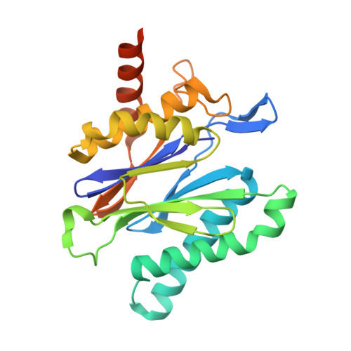
Proteasome subunit beta240Mycobacterium tuberculosisMutation(s): 0 
Gene Names: prcBMRA_2125
EC: 3.4.25.1
UniProt
Find proteins for P9WHT9 (Mycobacterium tuberculosis (strain ATCC 25618 / H37Rv))
Explore P9WHT9 
Go to UniProtKB:  P9WHT9
Entity Groups  
Sequence Clusters30% Identity50% Identity70% Identity90% Identity95% Identity100% Identity
UniProt GroupP9WHT9
Sequence Annotations
Expand
  • Reference Sequence

Find similar proteins by:  Sequence   |   3D Structure  

Entity ID: 3
MoleculeChains Sequence LengthOrganismDetailsImage
Fellutamide B3synthetic constructMutation(s): 0 
Sequence Annotations
Expand
  • Reference Sequence
Small Molecules
Ligands 1 Unique
IDChains Name / Formula / InChI Key2D Diagram3D Interactions
HXD
Query on HXD

Download Ideal Coordinates CCD File 
AB [auth k]
BB [auth l]
CB [auth m]
DB [auth n]
QA [auth a]
(3R)-3-HYDROXYDODECANOIC ACID
C12 H24 O3
MUCMKTPAZLSKTL-LLVKDONJSA-N
Experimental Data & Validation

Experimental Data

  • Method: X-RAY DIFFRACTION
  • Resolution: 2.50 Å
  • R-Value Free:  0.229 (Depositor), 0.220 (DCC) 
  • R-Value Work:  0.208 (Depositor), 0.200 (DCC) 
  • R-Value Observed: 0.208 (Depositor) 
Space Group: P 1 21 1
Unit Cell:
Length ( Å )Angle ( ˚ )
a = 170.186α = 90
b = 118.099β = 112.62
c = 194.347γ = 90
Software Package:
Software NamePurpose
CBASSdata collection
CNSrefinement
HKL-2000data reduction
HKL-2000data scaling
CNSphasing

Structure Validation

View Full Validation Report


Ligand Structure Quality Assessment 

Created with Raphaël 2.3.0Worse 01 BetterLigand structure goodness of fit to experimental dataBest fitted HXDClick on this verticalbar to view details

Entry History 

Deposition Data

  • Released Date: 2010-09-29 
  • Deposition Author(s): Li, D., Li, H.

Revision History  (Full details and data files)

  • Version 1.0: 2010-09-29
    Type: Initial release
  • Version 1.1: 2011-07-13
    Changes: Version format compliance
  • Version 2.0: 2024-03-27
    Changes: Advisory, Atomic model, Data collection, Database references, Derived calculations, Non-polymer description, Polymer sequence, Source and taxonomy, Structure summary
  • Version 2.1: 2024-11-06
    Changes: Structure summary