3BIN

Structure of the DAL-1 and TSLC1 (372-383) complex


Experimental Data Snapshot

  • Method: X-RAY DIFFRACTION
  • Resolution: 2.30 Å
  • R-Value Free: 0.230 
  • R-Value Work: 0.179 
  • R-Value Observed: 0.181 

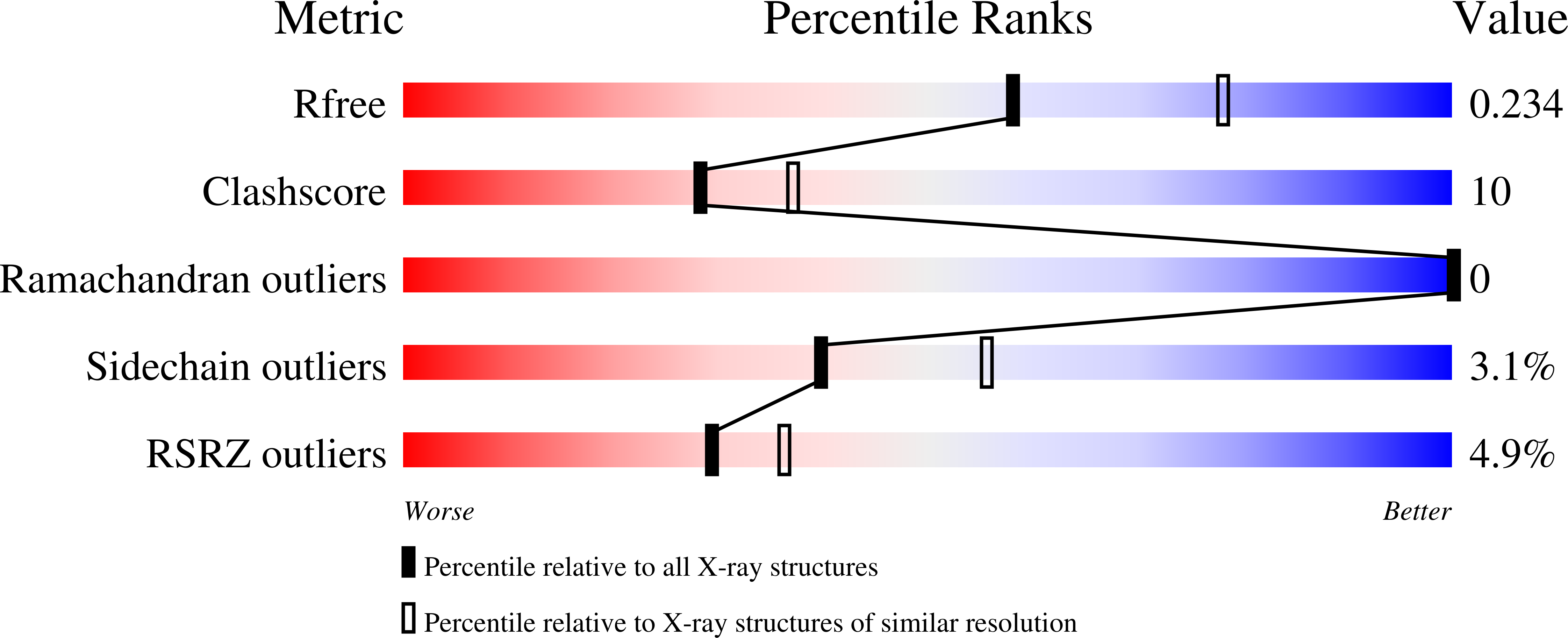
wwPDB Validation   3D Report Full Report


This is version 1.2 of the entry. See complete history


Literature

Structural basis of tumor suppressor in lung cancer 1 (TSLC1) binding to differentially expressed in adenocarcinoma of the lung (DAL-1/4.1B)

Busam, R.D.Thorsell, A.-G.Flores, A.Hammarstrom, M.Persson, C.Obrink, B.Hallberg, B.M.

(2011) J Biol Chem 286: 4511-4516

  • DOI: https://doi.org/10.1074/jbc.M110.174011
  • Primary Citation of Related Structures:  
    2HE7, 3BIN

  • PubMed Abstract: 

    Perturbed cell adhesion mechanisms are crucial for tumor invasion and metastasis. A cell adhesion protein, TSLC1 (tumor suppressor in lung cancer 1), is inactivated in a majority of metastatic cancers. DAL-1 (differentially expressed in adenocarcinoma of the lung protein), another tumor suppressor, binds through its FERM domain to the TSLC1 C-terminal, 4.1 glycophorin C-like, cytoplasmic domain. However, the molecular basis for this interaction is unknown. Here, we describe the crystal structure of a complex between the DAL-1 FERM domain and a portion of the TSLC1 cytoplasmic domain. DAL-1 binds to TSLC1 through conserved residues in a well defined hydrophobic pocket in the structural C-lobe of the DAL-1 FERM domain. From the crystal structure, it is apparent that Tyr(406) and Thr(408) in the TSLC1 cytoplasmic domain form the most important interactions with DAL-1, and this was also confirmed by surface plasmon resonance studies. Our results refute earlier exon deletion experiments that indicated that glycophorin C interacts with the α-lobe of 4.1 FERM domains.


  • Organizational Affiliation

    Structural Genomics Consortium, Department of Medical Biochemistry and Biophysics, Karolinska Institutet, SE-17177 Stockholm, Sweden.


Macromolecules
Find similar proteins by:  (by identity cutoff)  |  3D Structure
Entity ID: 1
MoleculeChains Sequence LengthOrganismDetailsImage
Band 4.1-like protein 3283Homo sapiensMutation(s): 0 
Gene Names: EPB41L3DAL1KIAA0987
UniProt & NIH Common Fund Data Resources
Find proteins for Q9Y2J2 (Homo sapiens)
Explore Q9Y2J2 
Go to UniProtKB:  Q9Y2J2
PHAROS:  Q9Y2J2
GTEx:  ENSG00000082397 
Entity Groups  
Sequence Clusters30% Identity50% Identity70% Identity90% Identity95% Identity100% Identity
UniProt GroupQ9Y2J2
Sequence Annotations
Expand
  • Reference Sequence

Find similar proteins by:  Sequence   |   3D Structure  

Entity ID: 2
MoleculeChains Sequence LengthOrganismDetailsImage
Cell adhesion molecule 112N/AMutation(s): 0 
UniProt & NIH Common Fund Data Resources
Find proteins for Q9BY67 (Homo sapiens)
Explore Q9BY67 
Go to UniProtKB:  Q9BY67
PHAROS:  Q9BY67
GTEx:  ENSG00000182985 
Entity Groups  
Sequence Clusters30% Identity50% Identity70% Identity90% Identity95% Identity100% Identity
UniProt GroupQ9BY67
Sequence Annotations
Expand
  • Reference Sequence
Experimental Data & Validation

Experimental Data

  • Method: X-RAY DIFFRACTION
  • Resolution: 2.30 Å
  • R-Value Free: 0.230 
  • R-Value Work: 0.179 
  • R-Value Observed: 0.181 
  • Space Group: P 65
Unit Cell:
Length ( Å )Angle ( ˚ )
a = 135α = 90
b = 135β = 90
c = 50.52γ = 120
Software Package:
Software NamePurpose
REFMACrefinement
PDB_EXTRACTdata extraction
MxCuBEdata collection
XDSdata reduction
XSCALEdata scaling

Structure Validation

View Full Validation Report


Entry History 

Revision History  (Full details and data files)

  • Version 1.0: 2008-01-15
    Type: Initial release
  • Version 1.1: 2011-07-13
    Changes: Advisory, Version format compliance
  • Version 1.2: 2024-03-13
    Changes: Data collection, Database references