3A4M | pdb_00003a4m

Crystal structure of archaeal O-phosphoseryl-tRNA(Sec) kinase


Experimental Data Snapshot

  • Method: X-RAY DIFFRACTION
  • Resolution: 1.79 Å
  • R-Value Free: 
    0.253 (Depositor), 0.250 (DCC) 
  • R-Value Work: 
    0.209 (Depositor), 0.200 (DCC) 
  • R-Value Observed: 
    0.211 (Depositor) 

wwPDB Validation   3D Report Full Report


Ligand Structure Quality Assessment 

Created with Raphaël 2.3.0Worse 01 BetterLigand structure goodness of fit to experimental dataBest fitted ADPClick on this verticalbar to view details

This is version 1.2 of the entry. See complete history


Literature

Structure of a tRNA-dependent kinase essential for selenocysteine decoding

Araiso, Y.Sherrer, R.L.Ishitani, R.Ho, J.M.L.Soll, D.Nureki, O.

(2009) Proc Natl Acad Sci U S A 106: 16215-16220

  • DOI: https://doi.org/10.1073/pnas.0908861106
  • Primary Citation of Related Structures:  
    3A4L, 3A4M, 3A4N

  • PubMed Abstract: 

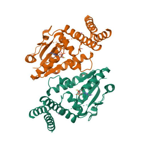
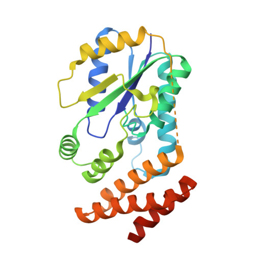
    Compared to bacteria, archaea and eukaryotes employ an additional enzyme for the biosynthesis of selenocysteine (Sec), the 21(st) natural amino acid (aa). An essential RNA-dependent kinase, O-phosphoseryl-tRNA(Sec) kinase (PSTK), converts seryl-tRNA(Sec) to O-phosphoseryl-tRNA(Sec), the immediate precursor of selenocysteinyl-tRNA(Sec). The sequence of Methanocaldococcus jannaschii PSTK (MjPSTK) suggests an N-terminal kinase domain (177 aa) followed by a presumed tRNA binding region (75 aa). The structures of MjPSTK complexed with ADP and AMPPNP revealed that this enzyme belongs to the P-loop kinase class, and that the kinase domain is closely related to gluconate kinase and adenylate kinase. ATP is bound by the P-loop domain (residues 11-18). Formed by antiparallel dimerization of two PSTK monomers, the enzyme structure shows a deep groove with positive electrostatic potential. Located in this groove is the enzyme's active site, which biochemical and genetic data suggest is composed of Asp-41, Arg-44, Glu-55, Tyr-82, Tyr-83, Met-86, and Met-132. Based on structural comparison with Escherichia coli adenylate kinase a docking model was generated that assigns these amino acids to the recognition of the terminal A76-Ser moieties of Ser-tRNA(Sec). The geometry and electrostatic environment of the groove in MjPSTK are perfectly complementary to the unusually long acceptor helix of tRNA(Sec).


  • Organizational Affiliation

    Department of Basic Medical Sciences, Institute of Medical Science, The University of Tokyo, 4-6-1 Shirokanedai, Minato-ku, Tokyo 108-8639, Japan.


Macromolecules
Find similar proteins by:  (by identity cutoff)  |  3D Structure
Entity ID: 1
MoleculeChains Sequence LengthOrganismDetailsImage
L-seryl-tRNA(Sec) kinase
A, B
260Methanocaldococcus jannaschiiMutation(s): 0 
Gene Names: MJ1538
EC: 2.7.1 (PDB Primary Data), 2.7.1.164 (UniProt)
UniProt
Find proteins for Q58933 (Methanocaldococcus jannaschii (strain ATCC 43067 / DSM 2661 / JAL-1 / JCM 10045 / NBRC 100440))
Explore Q58933 
Go to UniProtKB:  Q58933
Entity Groups  
Sequence Clusters30% Identity50% Identity70% Identity90% Identity95% Identity100% Identity
UniProt GroupQ58933
Sequence Annotations
Expand
  • Reference Sequence
Small Molecules
Ligands 4 Unique
IDChains Name / Formula / InChI Key2D Diagram3D Interactions
ADP
Query on ADP

Download Ideal Coordinates CCD File 
C [auth A],
G [auth B]
ADENOSINE-5'-DIPHOSPHATE
C10 H15 N5 O10 P2
XTWYTFMLZFPYCI-KQYNXXCUSA-N
IOD
Query on IOD

Download Ideal Coordinates CCD File 
E [auth A],
I [auth B]
IODIDE ION
I
XMBWDFGMSWQBCA-UHFFFAOYSA-M
EDO
Query on EDO

Download Ideal Coordinates CCD File 
F [auth A],
J [auth B],
K [auth B],
L [auth B]
1,2-ETHANEDIOL
C2 H6 O2
LYCAIKOWRPUZTN-UHFFFAOYSA-N
MG
Query on MG

Download Ideal Coordinates CCD File 
D [auth A],
H [auth B]
MAGNESIUM ION
Mg
JLVVSXFLKOJNIY-UHFFFAOYSA-N
Experimental Data & Validation

Experimental Data

  • Method: X-RAY DIFFRACTION
  • Resolution: 1.79 Å
  • R-Value Free:  0.253 (Depositor), 0.250 (DCC) 
  • R-Value Work:  0.209 (Depositor), 0.200 (DCC) 
  • R-Value Observed: 0.211 (Depositor) 
Space Group: P 1 21 1
Unit Cell:
Length ( Å )Angle ( ˚ )
a = 58.156α = 90
b = 74.524β = 113.37
c = 64.817γ = 90
Software Package:
Software NamePurpose
HKL-2000data collection
SHARPphasing
PHENIXrefinement
HKL-2000data reduction
HKL-2000data scaling

Structure Validation

View Full Validation Report


Ligand Structure Quality Assessment 

Created with Raphaël 2.3.0Worse 01 BetterLigand structure goodness of fit to experimental dataBest fitted ADPClick on this verticalbar to view details

Entry History 

Deposition Data

Revision History  (Full details and data files)

  • Version 1.0: 2009-10-20
    Type: Initial release
  • Version 1.1: 2011-07-13
    Changes: Version format compliance
  • Version 1.2: 2024-03-13
    Changes: Data collection, Database references, Derived calculations