2VMI

The structure of seleno-methionine labelled CBM51 from Clostridium perfringens GH95


Experimental Data Snapshot

  • Method: X-RAY DIFFRACTION
  • Resolution: 1.70 Å
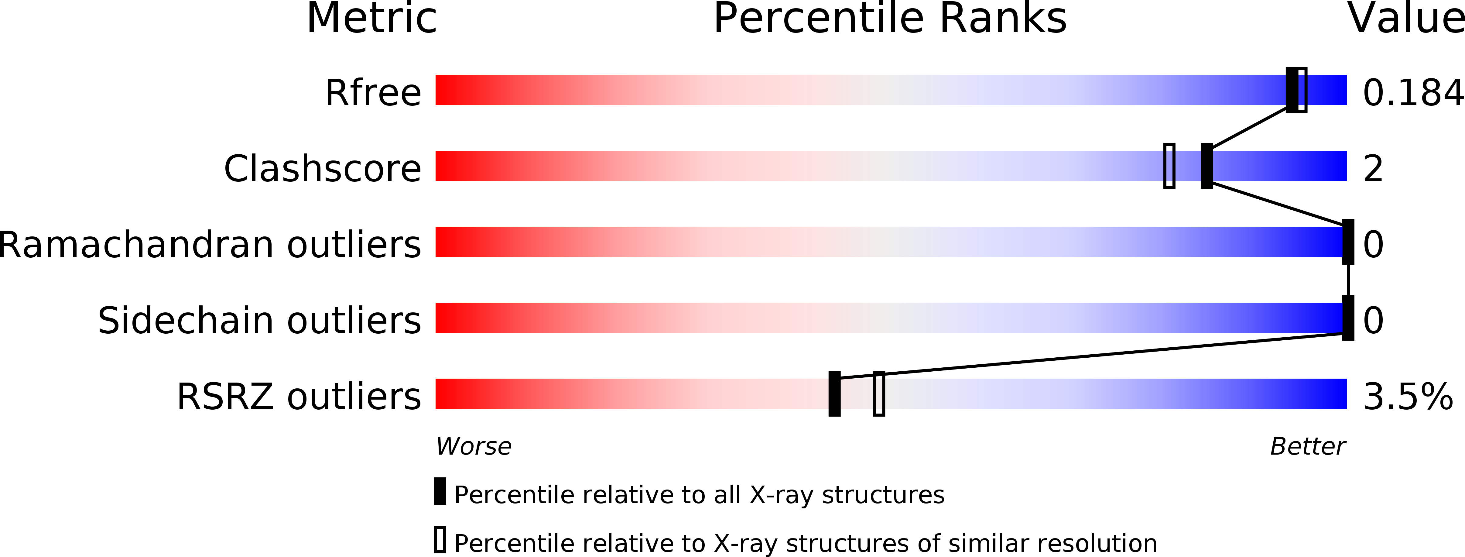
  • R-Value Free: 0.178 
  • R-Value Work: 0.150 
  • R-Value Observed: 0.151 

wwPDB Validation   3D Report Full Report


This is version 1.3 of the entry. See complete history


Literature

Divergent Modes of Glycan Recognition by a New Family of Carbohydrate-Binding Modules

Gregg, K.Finn, R.Abbott, D.W.Boraston, A.B.

(2008) J Biol Chem 283: 12604

  • DOI: https://doi.org/10.1074/jbc.M709865200
  • Primary Citation of Related Structures:  
    2VMG, 2VMH, 2VMI, 2VNG, 2VNO, 2VNR

  • PubMed Abstract: 

    The genomes of myonecrotic Clostridium perfringens isolates contain genes encoding a large and fascinating array of highly modular glycoside hydrolase enzymes. Although the catalytic activities of many of these enzymes are somewhat predictable based on their amino acid sequences, the functions of their abundant ancillary modules are not and remain poorly studied. Here, we present the structural and functional analysis of a new family of ancillary carbohydrate-binding modules (CBMs), CBM51, which was previously annotated in data bases as the novel putative CBM domain. The high resolution crystal structures of two CBM51 members, GH95CBM51 and GH98CBM51, from a putative family 95 alpha-fucosidase and from a family 98 blood group A/B antigen-specific endo-beta-galactosidase, respectively, showed them to have highly similar beta-sandwich folds. However, GH95CBM51 was shown by glycan microarray screening, isothermal titration calorimetry, and x-ray crystallography to bind galactose residues, whereas the same analyses of GH98CBM51 revealed specificity for the blood group A/B antigens through non-conserved interactions. Overall, this work identifies a new family of CBMs with many members having apparent specificity for eukaryotic glycans, in keeping with the glycan-rich environment C. perfringens would experience in its host. However, a wider bioinformatic analysis of this CBM family also indicated a large number of members in non-pathogenic environmental bacteria, suggesting a role in the recognition of environmental glycans.


  • Organizational Affiliation

    Department of Biochemistry and Microbiology, University of Victoria, Victoria, British Columbia V8W 3P6, Canada.


Macromolecules
Find similar proteins by:  (by identity cutoff)  |  3D Structure
Entity ID: 1
MoleculeChains Sequence LengthOrganismDetailsImage
FIBRONECTIN TYPE III DOMAIN PROTEIN151Clostridium perfringensMutation(s): 0 
UniProt
Find proteins for A0A0H2YQB3 (Clostridium perfringens (strain ATCC 13124 / DSM 756 / JCM 1290 / NCIMB 6125 / NCTC 8237 / Type A))
Explore A0A0H2YQB3 
Go to UniProtKB:  A0A0H2YQB3
Entity Groups  
Sequence Clusters30% Identity50% Identity70% Identity90% Identity95% Identity100% Identity
UniProt GroupA0A0H2YQB3
Sequence Annotations
Expand
  • Reference Sequence
Small Molecules
Ligands 1 Unique
IDChains Name / Formula / InChI Key2D Diagram3D Interactions
CA
Query on CA

Download Ideal Coordinates CCD File 
B [auth A]CALCIUM ION
Ca
BHPQYMZQTOCNFJ-UHFFFAOYSA-N
Modified Residues  1 Unique
IDChains TypeFormula2D DiagramParent
MSE
Query on MSE
A
L-PEPTIDE LINKINGC5 H11 N O2 SeMET
Experimental Data & Validation

Experimental Data

  • Method: X-RAY DIFFRACTION
  • Resolution: 1.70 Å
  • R-Value Free: 0.178 
  • R-Value Work: 0.150 
  • R-Value Observed: 0.151 
  • Space Group: P 65
Unit Cell:
Length ( Å )Angle ( ˚ )
a = 76.721α = 90
b = 76.721β = 90
c = 51.771γ = 120
Software Package:
Software NamePurpose
REFMACrefinement

Structure Validation

View Full Validation Report


Entry History 

Deposition Data

Revision History  (Full details and data files)

  • Version 1.0: 2008-02-19
    Type: Initial release
  • Version 1.1: 2011-05-08
    Changes: Version format compliance
  • Version 1.2: 2011-07-13
    Changes: Version format compliance
  • Version 1.3: 2024-11-06
    Changes: Data collection, Database references, Derived calculations, Other, Structure summary