2JGV | pdb_00002jgv

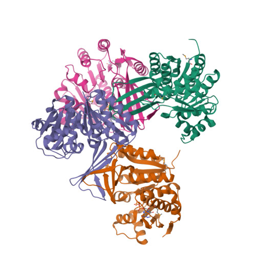
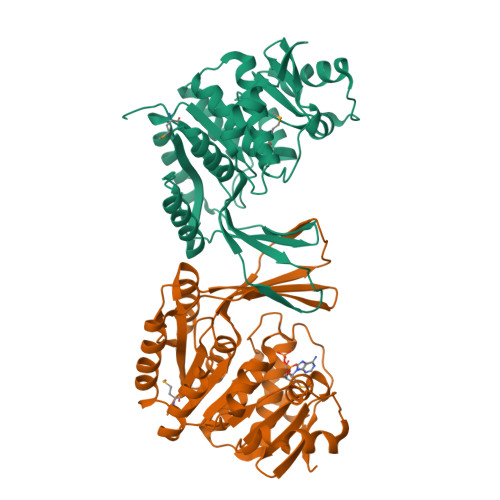
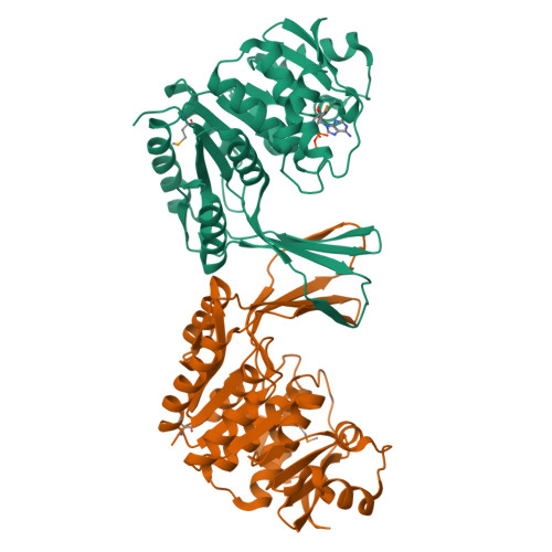
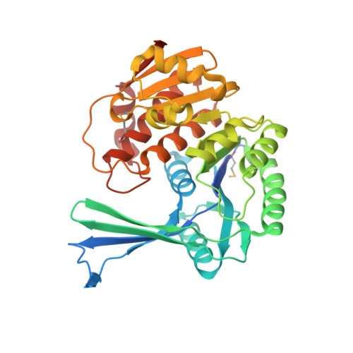
STRUCTURE OF Staphylococcus aureus D-TAGATOSE-6-PHOSPHATE KINASE in complex with ADP


Experimental Data Snapshot

  • Method: X-RAY DIFFRACTION
  • Resolution: 2.00 Å
  • R-Value Free: 
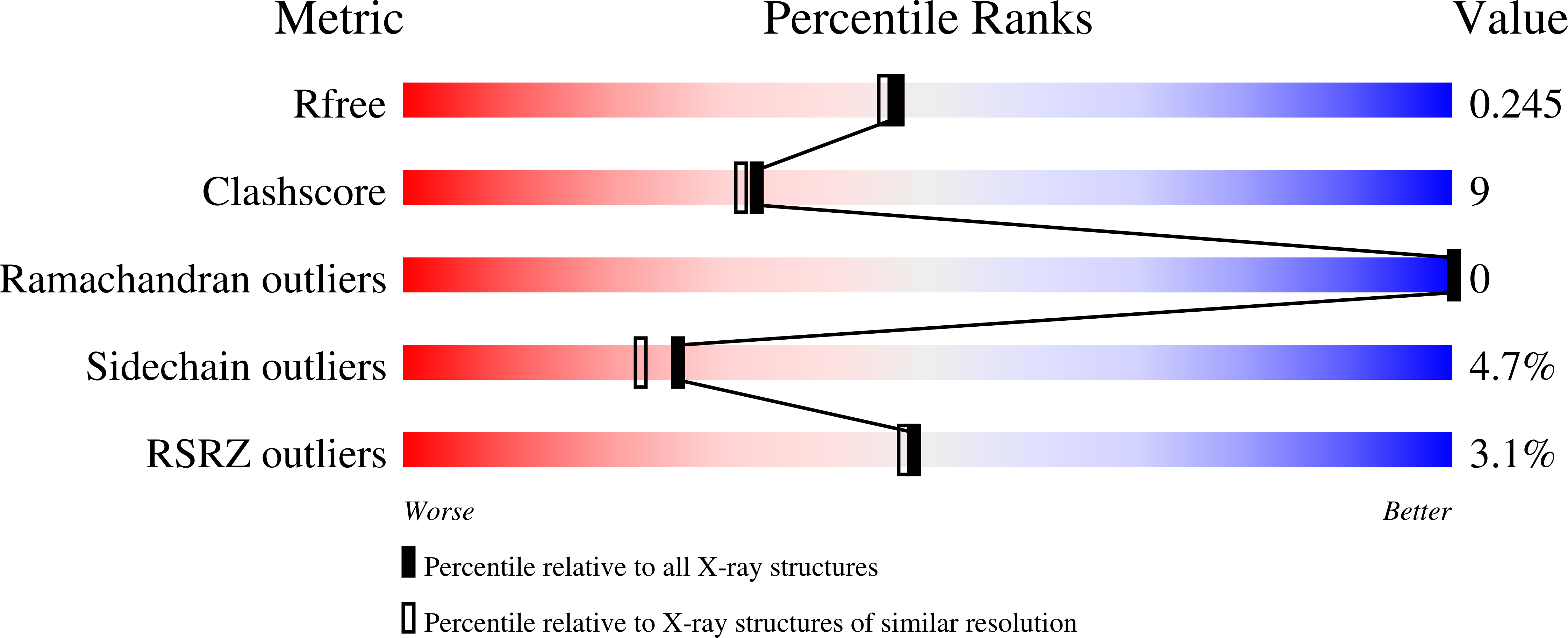
    0.247 (Depositor), 0.240 (DCC) 
  • R-Value Work: 
    0.198 (Depositor), 0.200 (DCC) 
  • R-Value Observed: 
    0.201 (Depositor) 

Starting Model: experimental
View more details

wwPDB Validation   3D Report Full Report


Ligand Structure Quality Assessment 

Created with Raphaël 2.3.0Worse 01 BetterLigand structure goodness of fit to experimental dataBest fitted ADPClick on this verticalbar to view details

This is version 1.4 of the entry. See complete history


Literature

Structures of Staphylococcus Aureus D-Tagatose-6-Phosphate Kinase Implicate Domain Motions in Specificity and Mechanism.

Miallau, L.Hunter, W.N.Mcsweeney, S.M.Leonard, G.A.

(2007) J Biological Chem 282: 19948

  • DOI: https://doi.org/10.1074/jbc.M701480200
  • Primary Citation of Related Structures:  
    2JG1, 2JGV

  • PubMed Abstract: 

    High resolution structures of Staphylococcus aureus d-tagatose-6-phosphate kinase (LacC) in two crystal forms are herein reported. The structures define LacC in apoform, in binary complexes with ADP or the co-factor analogue AMP-PNP, and in a ternary complex with AMP-PNP and D-tagatose-6-phosphate. The tertiary structure of the LacC monomer, which is closely related to other members of the pfkB subfamily of carbohydrate kinases, is composed of a large alpha/beta core domain and a smaller, largely beta "lid." Four extended polypeptide segments connect these two domains. Dimerization of LacC occurs via interactions between lid domains, which come together to form a beta-clasp structure. Residues from both subunits contribute to substrate binding. LacC adopts a closed structure required for phosphoryl transfer only when both substrate and co-factor are bound. A reaction mechanism similar to that used by other phosphoryl transferases is proposed, although unusually, when both substrate and co-factor are bound to the enzyme two Mg(2+) ions are observed in the active site. A new motif of amino acid sequence conservation common to the pfkB subfamily of carbohydrate kinases is identified.


  • Organizational Affiliation

    Macromolecular Crystallography Group, European Synchrotron Radiation Facility, 38043 Grenoble, France.


Macromolecules
Find similar proteins by:  (by identity cutoff)  |  3D Structure
Entity ID: 1
MoleculeChains Sequence LengthOrganismDetailsImage
TAGATOSE-6-PHOSPHATE KINASE
A, B, C, D
330Staphylococcus aureus subsp. aureus NCTC 8325Mutation(s): 0 
EC: 2.7.1.144
UniProt
Find proteins for P0A0B9 (Staphylococcus aureus (strain NCTC 8325 / PS 47))
Explore P0A0B9 
Go to UniProtKB:  P0A0B9
Entity Groups  
Sequence Clusters30% Identity50% Identity70% Identity90% Identity95% Identity100% Identity
UniProt GroupP0A0B9
Sequence Annotations
Expand
  • Reference Sequence
Small Molecules
Modified Residues  1 Unique
IDChains TypeFormula2D DiagramParent
MSE
Query on MSE
A, B, C, D
L-PEPTIDE LINKINGC5 H11 N O2 SeMET
Experimental Data & Validation

Experimental Data

  • Method: X-RAY DIFFRACTION
  • Resolution: 2.00 Å
  • R-Value Free:  0.247 (Depositor), 0.240 (DCC) 
  • R-Value Work:  0.198 (Depositor), 0.200 (DCC) 
  • R-Value Observed: 0.201 (Depositor) 
Space Group: P 1 21 1
Unit Cell:
Length ( Å )Angle ( ˚ )
a = 77.548α = 90
b = 96.38β = 94.29
c = 94.841γ = 90
Software Package:
Software NamePurpose
REFMACrefinement
MOSFLMdata reduction
SCALAdata scaling
PHASERphasing

Structure Validation

View Full Validation Report


Ligand Structure Quality Assessment 

Created with Raphaël 2.3.0Worse 01 BetterLigand structure goodness of fit to experimental dataBest fitted ADPClick on this verticalbar to view details

Entry History 

Deposition Data

Revision History  (Full details and data files)

  • Version 1.0: 2007-04-24
    Type: Initial release
  • Version 1.1: 2011-05-08
    Changes: Version format compliance
  • Version 1.2: 2011-07-13
    Changes: Version format compliance
  • Version 1.3: 2023-12-13
    Changes: Data collection, Database references, Derived calculations, Other, Refinement description
  • Version 1.4: 2024-10-16
    Changes: Structure summary