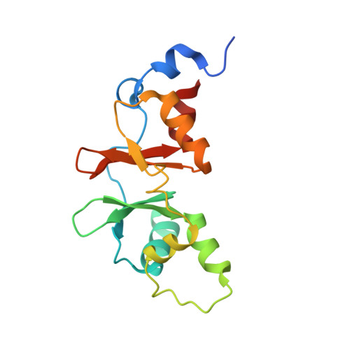
2UV7

Crystal Structure of a CBS domain pair from the regulatory gamma1 subunit of human AMPK in complex with AMP


Experimental Data Snapshot

  • Method: X-RAY DIFFRACTION
  • Resolution: 2.00 Å
  • R-Value Free: 0.230 
  • R-Value Work: 0.158 
  • R-Value Observed: 0.162 

wwPDB Validation   3D Report Full Report


This is version 1.3 of the entry. See complete history


Literature

Structure of a Cbs-Domain Pair from the Regulatory Gamma1 Subunit of Human Ampk in Complex with AMP and Zmp.

Day, P.Sharff, A.Parra, L.Cleasby, A.Williams, M.Horer, S.Nar, H.Redemann, N.Tickle, I.Yon, J.

(2007) Acta Crystallogr D Biol Crystallogr 63: 587

  • DOI: https://doi.org/10.1107/S0907444907009110
  • Primary Citation of Related Structures:  
    2UV4, 2UV5, 2UV6, 2UV7

  • PubMed Abstract: 

    AMP-activated kinase (AMPK) is central to sensing energy status in eukaryotic cells via binding of AMP and ATP to CBS (cystathionine beta-synthase) domains in the regulatory gamma subunit. The structure of a CBS-domain pair from human AMPK gamma1 in complex with the physiological activator AMP and the pharmacological activator ZMP (AICAR) is presented.


  • Organizational Affiliation

    Astex Therapeutics Ltd, 436 Cambridge Science Park, Milton Road, Cambridge CB4 0QA, England. [email protected]


Macromolecules
Find similar proteins by:  (by identity cutoff)  |  3D Structure
Entity ID: 1
MoleculeChains Sequence LengthOrganismDetailsImage
5'-AMP-ACTIVATED PROTEIN KINASE SUBUNIT GAMMA-1152Homo sapiensMutation(s): 1 
UniProt & NIH Common Fund Data Resources
Find proteins for P54619 (Homo sapiens)
Explore P54619 
Go to UniProtKB:  P54619
PHAROS:  P54619
GTEx:  ENSG00000181929 
Entity Groups  
Sequence Clusters30% Identity50% Identity70% Identity90% Identity95% Identity100% Identity
UniProt GroupP54619
Sequence Annotations
Expand
  • Reference Sequence
Small Molecules
Ligands 1 Unique
IDChains Name / Formula / InChI Key2D Diagram3D Interactions
AMP
Query on AMP

Download Ideal Coordinates CCD File 
B [auth A]ADENOSINE MONOPHOSPHATE
C10 H14 N5 O7 P
UDMBCSSLTHHNCD-KQYNXXCUSA-N
Binding Affinity Annotations 
IDSourceBinding Affinity
AMP BindingDB:  2UV7 Kd: 3700 (nM) from 1 assay(s)
EC50: min: 1400, max: 1519 (nM) from 2 assay(s)
Experimental Data & Validation

Experimental Data

  • Method: X-RAY DIFFRACTION
  • Resolution: 2.00 Å
  • R-Value Free: 0.230 
  • R-Value Work: 0.158 
  • R-Value Observed: 0.162 
  • Space Group: C 1 2 1
Unit Cell:
Length ( Å )Angle ( ˚ )
a = 65.995α = 90
b = 43.836β = 118.79
c = 55.734γ = 90
Software Package:
Software NamePurpose
REFMACrefinement
d*TREKdata reduction
d*TREKdata scaling

Structure Validation

View Full Validation Report


Entry History 

Deposition Data

Revision History  (Full details and data files)

  • Version 1.0: 2007-05-08
    Type: Initial release
  • Version 1.1: 2011-05-08
    Changes: Version format compliance
  • Version 1.2: 2011-07-13
    Changes: Version format compliance
  • Version 1.3: 2024-05-08
    Changes: Data collection, Database references, Other