2DD4 | pdb_00002dd4

Thiocyanate hydrolase (SCNase) from Thiobacillus thioparus recombinant apo-enzyme


Experimental Data Snapshot

  • Method: X-RAY DIFFRACTION
  • Resolution: 2.06 Å
  • R-Value Free: 
    0.193 (Depositor), 0.170 (DCC) 
  • R-Value Work: 
    0.165 (Depositor), 0.160 (DCC) 
  • R-Value Observed: 
    0.165 (Depositor) 

wwPDB Validation   3D Report Full Report


Ligand Structure Quality Assessment 

Created with Raphaël 2.3.0Worse 01 BetterLigand structure goodness of fit to experimental dataBest fitted FRUClick on this verticalbar to view details

This is version 2.2 of the entry. See complete history


Literature

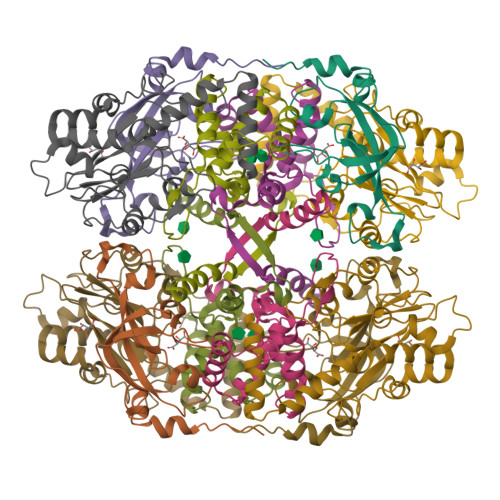
Structure of thiocyanate hydrolase: a new nitrile hydratase family protein with a novel five-coordinate cobalt(III) center.

Arakawa, T.Kawano, Y.Kataoka, S.Katayama, Y.Kamiya, N.Yohda, M.Odaka, M.

(2007) J Mol Biology 366: 1497-1509

  • DOI: https://doi.org/10.1016/j.jmb.2006.12.011
  • Primary Citation of Related Structures:  
    2DD4, 2DD5

  • PubMed Abstract: 

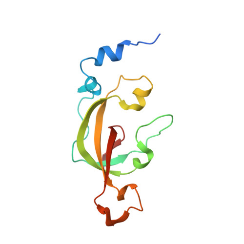
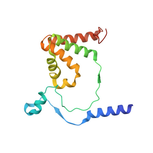
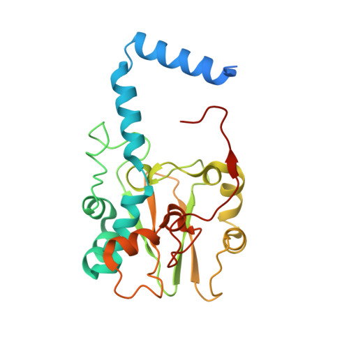
    Thiocyanate hydrolase (SCNase) of Thiobacillus thioparus THI115 is a cobalt(III)-containing enzyme catalyzing the degradation of thiocyanate to carbonyl sulfide and ammonia. We determined the crystal structures of the apo- and native SCNases at a resolution of 2.0 A. SCNases in both forms had a conserved hetero-dodecameric structure, (alphabetagamma)(4). Four alphabetagamma hetero-trimers were structurally equivalent. One alphabetagamma hetero-trimer was composed of the core domain and the betaN domain, which was located at the center of the molecule and linked the hetero-trimers with novel quaternary interfaces. In both the apo- and native SCNases, the core domain was structurally conserved between those of iron and cobalt-types of nitrile hydratase (NHase). Native SCNase possessed the post-translationally modified cysteine ligands, gammaCys131-SO(2)H and gammaCys133-SOH like NHases. However, the low-spin cobalt(III) was found to be in the distorted square-pyramidal geometry, which had not been reported before in any protein. The size as well as the electrostatic properties of the substrate-binding pocket was totally different from NHases with respect to the charge distribution and the substrate accessibility, which rationally explains the differences in the substrate preference between SCNase and NHase.


  • Organizational Affiliation

    Department of Biotechnology and Life Science, Graduate School of Technology, Tokyo University of Agriculture and Technology, 2-24-16 Naka-cho, Koganei, Tokyo 184-8588, Japan.


Macromolecules
Find similar proteins by:  (by identity cutoff)  |  3D Structure
Entity ID: 1
MoleculeChains Sequence LengthOrganismDetailsImage
Thiocyanate hydrolase alpha subunit
A, D, G, J
126Thiobacillus thioparusMutation(s): 0 
EC: 3.5.5.8
UniProt
Find proteins for O66187 (Thiobacillus thioparus)
Explore O66187 
Go to UniProtKB:  O66187
Entity Groups  
Sequence Clusters30% Identity50% Identity70% Identity90% Identity95% Identity100% Identity
UniProt GroupO66187
Sequence Annotations
Expand
  • Reference Sequence
Find similar proteins by:  (by identity cutoff)  |  3D Structure
Entity ID: 2
MoleculeChains Sequence LengthOrganismDetailsImage
Thiocyanate hydrolase beta subunit
B, E, H, K
157Thiobacillus thioparusMutation(s): 0 
EC: 3.5.5.8
UniProt
Find proteins for O66186 (Thiobacillus thioparus)
Explore O66186 
Go to UniProtKB:  O66186
Entity Groups  
Sequence Clusters30% Identity50% Identity70% Identity90% Identity95% Identity100% Identity
UniProt GroupO66186
Sequence Annotations
Expand
  • Reference Sequence
Find similar proteins by:  (by identity cutoff)  |  3D Structure
Entity ID: 3
MoleculeChains Sequence LengthOrganismDetailsImage
Thiocyanate hydrolase gamma subunit
C, F, I, L
243Thiobacillus thioparusMutation(s): 0 
EC: 3.5.5.8
UniProt
Find proteins for O66188 (Thiobacillus thioparus)
Explore O66188 
Go to UniProtKB:  O66188
Entity Groups  
Sequence Clusters30% Identity50% Identity70% Identity90% Identity95% Identity100% Identity
UniProt GroupO66188
Sequence Annotations
Expand
  • Reference Sequence
Small Molecules
Ligands 2 Unique
IDChains Name / Formula / InChI Key2D Diagram3D Interactions
FRU
Query on FRU

Download Ideal Coordinates CCD File 
M [auth B]
N [auth B]
O [auth C]
T [auth H]
U [auth H]
beta-D-fructofuranose
C6 H12 O6
RFSUNEUAIZKAJO-ARQDHWQXSA-N
TLA
Query on TLA

Download Ideal Coordinates CCD File 
AA [auth L]
BA [auth L]
P [auth C]
Q [auth C]
R [auth F]
L(+)-TARTARIC ACID
C4 H6 O6
FEWJPZIEWOKRBE-JCYAYHJZSA-N
Experimental Data & Validation

Experimental Data

  • Method: X-RAY DIFFRACTION
  • Resolution: 2.06 Å
  • R-Value Free:  0.193 (Depositor), 0.170 (DCC) 
  • R-Value Work:  0.165 (Depositor), 0.160 (DCC) 
  • R-Value Observed: 0.165 (Depositor) 
Space Group: P 21 21 21
Unit Cell:
Length ( Å )Angle ( ˚ )
a = 170.307α = 90
b = 175.602β = 90
c = 114.791γ = 90
Software Package:
Software NamePurpose
CNSrefinement
HKL-2000data reduction
SCALEPACKdata scaling
SOLVEphasing

Structure Validation

View Full Validation Report


Ligand Structure Quality Assessment 

Created with Raphaël 2.3.0Worse 01 BetterLigand structure goodness of fit to experimental dataBest fitted FRUClick on this verticalbar to view details

Entry History 

Deposition Data

Revision History  (Full details and data files)

  • Version 1.0: 2007-01-30
    Type: Initial release
  • Version 1.1: 2008-04-30
    Changes: Version format compliance
  • Version 1.2: 2011-07-13
    Changes: Version format compliance
  • Version 2.0: 2019-07-10
    Changes: Atomic model, Data collection, Derived calculations, Non-polymer description, Structure summary
  • Version 2.1: 2020-07-29
    Type: Remediation
    Reason: Carbohydrate remediation
    Changes: Data collection, Database references, Derived calculations, Structure summary
  • Version 2.2: 2024-03-13
    Changes: Data collection, Database references, Structure summary