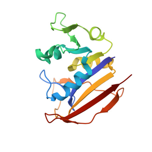
2CIG

Dihydrofolate reductase from Mycobacterium tuberculosis inhibited by the acyclic 4R isomer of INH-NADP a derivative of the prodrug isoniazid.


Experimental Data Snapshot

  • Method: X-RAY DIFFRACTION
  • Resolution: 1.90 Å
  • R-Value Free: 0.212 
  • R-Value Work: 0.178 
  • R-Value Observed: 0.180 

Starting Model: experimental
View more details

wwPDB Validation   3D Report Full Report


Ligand Structure Quality Assessment 


This is version 1.4 of the entry. See complete history


Literature

Mycobacterium Tuberculosis Dihydrofolate Reductase is a Target for Isoniazid

Argyrou, A.Vetting, M.W.Aladegbami, B.Blanchard, J.S.

(2006) Nat Struct Mol Biol 13: 408-413

  • DOI: https://doi.org/10.1038/nsmb1089
  • Primary Citation of Related Structures:  
    2CIG

  • PubMed Abstract: 

    Isoniazid is a key drug used in the treatment of tuberculosis. Isoniazid is a pro-drug, which, after activation by the katG-encoded catalase peroxidase, reacts nonenzymatically with NAD(+) and NADP(+) to generate several isonicotinoyl adducts of these pyridine nucleotides. One of these, the acyclic 4S isomer of isoniazid-NAD, targets the inhA-encoded enoyl-ACP reductase, an enzyme essential for mycolic acid biosynthesis in Mycobacterium tuberculosis. Here we show that the acyclic 4R isomer of isoniazid-NADP inhibits the M. tuberculosis dihydrofolate reductase (DHFR), an enzyme essential for nucleic acid synthesis. This biologically relevant form of the isoniazid adduct is a subnanomolar bisubstrate inhibitor of M. tuberculosis DHFR. Expression of M. tuberculosis DHFR in Mycobacterium smegmatis mc(2)155 protects cells against growth inhibition by isoniazid by sequestering the drug. Thus, M. tuberculosis DHFR is the first new target for isoniazid identified in the last decade.


  • Organizational Affiliation

    Department of Biochemistry, Albert Einstein College of Medicine, 1300 Morris Park Avenue, Bronx, New York 10461, USA.


Macromolecules
Find similar proteins by:  (by identity cutoff)  |  3D Structure
Entity ID: 1
MoleculeChains Sequence LengthOrganismDetailsImage
DIHYDROFOLATE REDUCTASE159Mycobacterium tuberculosis H37RvMutation(s): 0 
EC: 1.5.1.3
UniProt
Find proteins for P9WNX1 (Mycobacterium tuberculosis (strain ATCC 25618 / H37Rv))
Explore P9WNX1 
Go to UniProtKB:  P9WNX1
Entity Groups  
Sequence Clusters30% Identity50% Identity70% Identity90% Identity95% Identity100% Identity
UniProt GroupP9WNX1
Sequence Annotations
Expand
  • Reference Sequence
Small Molecules
Ligands 3 Unique
IDChains Name / Formula / InChI Key2D Diagram3D Interactions
1DG
Query on 1DG

Download Ideal Coordinates CCD File 
B [auth A](4R)-ISONICOTINIC-ACETYL-NICOTINAMIDE-ADENINE DINUCLEOTIDE
C27 H33 N8 O18 P3
FPZCWRGJBUDVSB-MXUAFQDZSA-N
SO4
Query on SO4

Download Ideal Coordinates CCD File 
G [auth A],
H [auth A]
SULFATE ION
O4 S
QAOWNCQODCNURD-UHFFFAOYSA-L
GOL
Query on GOL

Download Ideal Coordinates CCD File 
C [auth A],
D [auth A],
E [auth A],
F [auth A]
GLYCEROL
C3 H8 O3
PEDCQBHIVMGVHV-UHFFFAOYSA-N
Experimental Data & Validation

Experimental Data

  • Method: X-RAY DIFFRACTION
  • Resolution: 1.90 Å
  • R-Value Free: 0.212 
  • R-Value Work: 0.178 
  • R-Value Observed: 0.180 
  • Space Group: P 41
Unit Cell:
Length ( Å )Angle ( ˚ )
a = 60.67α = 90
b = 60.67β = 90
c = 59.147γ = 90
Software Package:
Software NamePurpose
REFMACrefinement
MOSFLMdata reduction
CCP4data scaling
AMoREphasing

Structure Validation

View Full Validation Report


Ligand Structure Quality Assessment 


Entry History 

Deposition Data

Revision History  (Full details and data files)

  • Version 1.0: 2006-04-25
    Type: Initial release
  • Version 1.1: 2011-05-08
    Changes: Version format compliance
  • Version 1.2: 2011-07-13
    Changes: Version format compliance
  • Version 1.3: 2018-06-20
    Changes: Data collection, Database references
  • Version 1.4: 2023-12-13
    Changes: Data collection, Database references, Derived calculations, Other, Refinement description