2A0F

Structure of D236A mutant E. coli Aspartate Transcarbamoylase in presence of Phosphonoacetamide at 2.90 A resolution


Experimental Data Snapshot

  • Method: X-RAY DIFFRACTION
  • Resolution: 2.90 Å
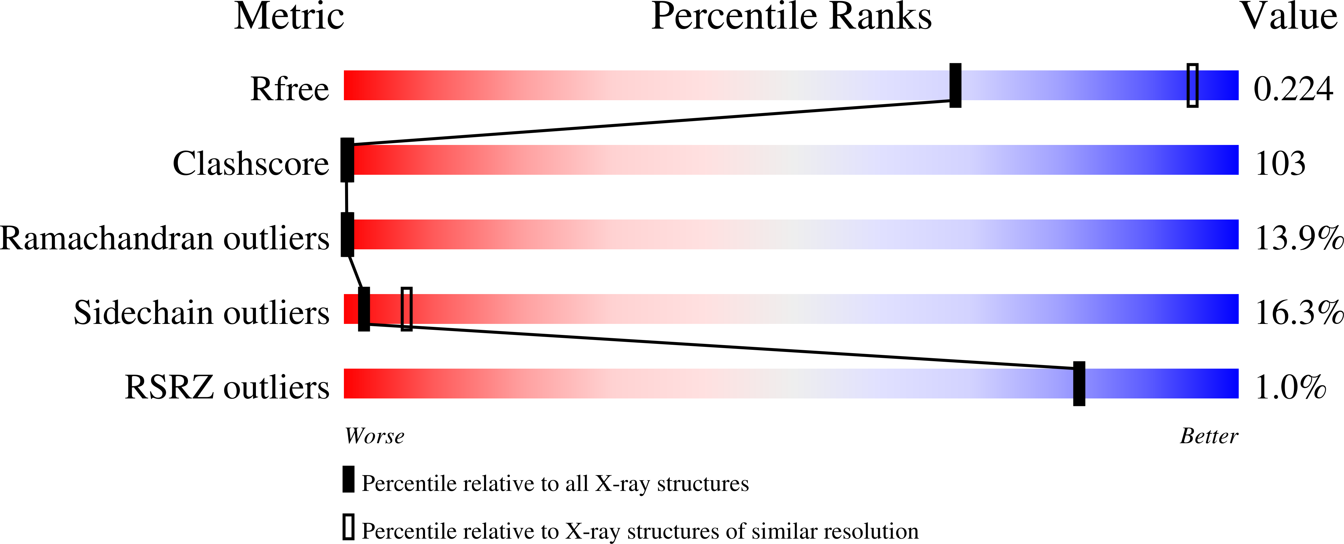
  • R-Value Free: 0.279 
  • R-Value Work: 0.217 
  • R-Value Observed: 0.217 

Starting Model: experimental
View more details

wwPDB Validation   3D Report Full Report


This is version 1.6 of the entry. See complete history


Literature

Structure of the E.coli Aspartate Transcarbamoylase Trapped in the Middle of the Catalytic Cycle.

Stieglitz, K.A.Dusinberre, K.J.Cardia, J.P.Tsuruta, H.Kantrowitz, E.R.

(2005) J Mol Biol 352: 478-486

  • DOI: https://doi.org/10.1016/j.jmb.2005.07.046
  • Primary Citation of Related Structures:  
    2A0F

  • PubMed Abstract: 

    Snapshots of the catalytic cycle of the allosteric enzyme aspartate transcarbamoylase have been obtained via X-ray crystallography. The enzyme in the high-activity high-affinity R state contains two catalytic chains in the asymmetric unit that are different. The active site in one chain is empty, while the active site in the other chain contains an analog of the first substrate to bind in the ordered mechanism of the reaction. Small angle X-ray scattering shows that once the enzyme is converted to the R state, by substrate binding, the enzyme remains in the R state until substrates are exhausted. Thus, this structure represents the active form of the enzyme trapped at two different stages in the catalytic cycle, before the substrates bind (or after the products are released), and after the first substrate binds. Opening and closing of the catalytic chain domains explains how the catalytic cycle occurs while the enzyme remains globally in the R-quaternary structure.


  • Organizational Affiliation

    Department of Chemistry, Merkert Chemistry Center, Boston College, Chestnut Hill, MA 02467, USA.


Macromolecules
Find similar proteins by:  (by identity cutoff)  |  3D Structure
Entity ID: 1
MoleculeChains Sequence LengthOrganismDetailsImage
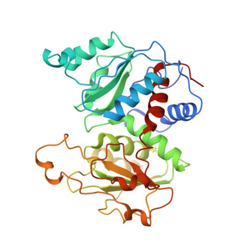
Aspartate carbamoyltransferase catalytic chain
A, C
310Escherichia coliMutation(s): 1 
Gene Names: pyrB
EC: 2.1.3.2
UniProt
Find proteins for P0A786 (Escherichia coli (strain K12))
Explore P0A786 
Go to UniProtKB:  P0A786
Entity Groups  
Sequence Clusters30% Identity50% Identity70% Identity90% Identity95% Identity100% Identity
UniProt GroupP0A786
Sequence Annotations
Expand
  • Reference Sequence
Find similar proteins by:  (by identity cutoff)  |  3D Structure
Entity ID: 2
MoleculeChains Sequence LengthOrganismDetailsImage
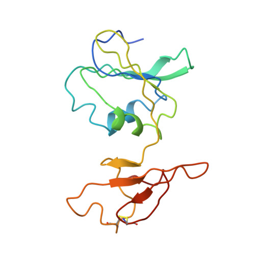
Aspartate carbamoyltransferase regulatory chain
B, D
153Escherichia coliMutation(s): 0 
Gene Names: pyrI
UniProt
Find proteins for P0A7F3 (Escherichia coli (strain K12))
Explore P0A7F3 
Go to UniProtKB:  P0A7F3
Entity Groups  
Sequence Clusters30% Identity50% Identity70% Identity90% Identity95% Identity100% Identity
UniProt GroupP0A7F3
Sequence Annotations
Expand
  • Reference Sequence
Experimental Data & Validation

Experimental Data

  • Method: X-RAY DIFFRACTION
  • Resolution: 2.90 Å
  • R-Value Free: 0.279 
  • R-Value Work: 0.217 
  • R-Value Observed: 0.217 
  • Space Group: P 3 2 1
Unit Cell:
Length ( Å )Angle ( ˚ )
a = 120.452α = 90
b = 120.452β = 90
c = 155.242γ = 120
Software Package:
Software NamePurpose
CNSrefinement
CNSphasing

Structure Validation

View Full Validation Report


Entry History 

Deposition Data

Revision History  (Full details and data files)

  • Version 1.0: 2005-09-27
    Type: Initial release
  • Version 1.1: 2008-04-30
    Changes: Version format compliance
  • Version 1.2: 2011-07-13
    Changes: Version format compliance
  • Version 1.3: 2017-10-11
    Changes: Refinement description
  • Version 1.4: 2021-10-20
    Changes: Database references, Derived calculations
  • Version 1.5: 2023-08-23
    Changes: Data collection, Refinement description
  • Version 1.6: 2024-11-13
    Changes: Structure summary