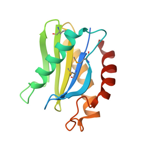
1IFQ

Sec22b N-terminal domain


Experimental Data Snapshot

  • Method: X-RAY DIFFRACTION
  • Resolution: 2.40 Å
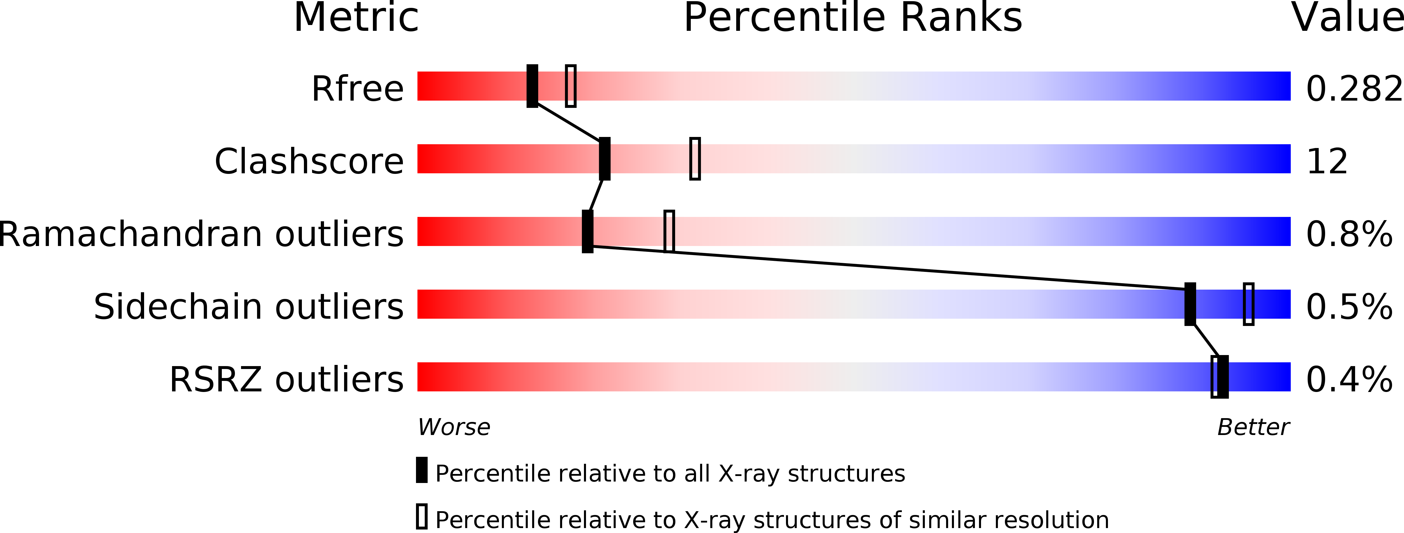
  • R-Value Free: 0.274 
  • R-Value Work: 0.231 

wwPDB Validation   3D Report Full Report


This is version 1.3 of the entry. See complete history


Literature

A novel snare N-terminal domain revealed by the crystal structure of Sec22b.

Gonzalez Jr., L.C.Weis, W.I.Scheller, R.H.

(2001) J Biol Chem 276: 24203-24211

  • DOI: https://doi.org/10.1074/jbc.M101584200
  • Primary Citation of Related Structures:  
    1IFQ

  • PubMed Abstract: 

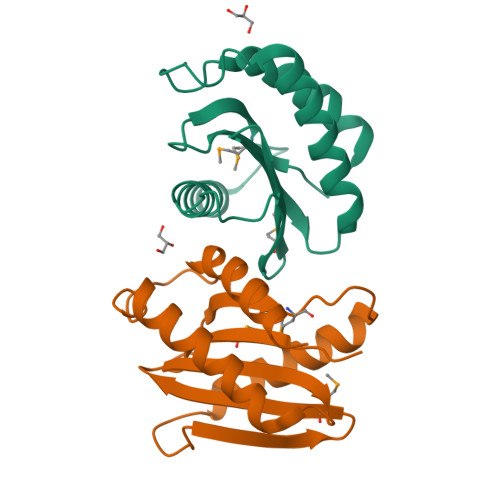
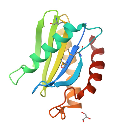
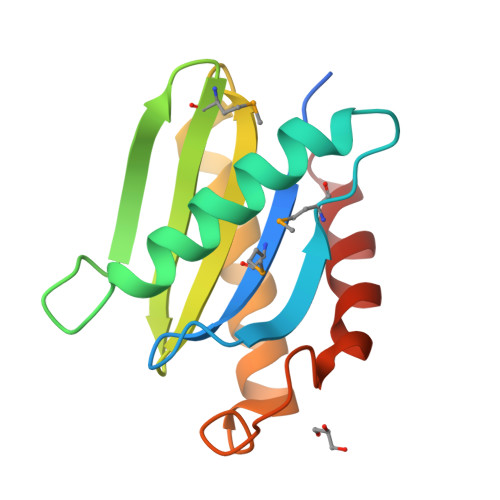
    Intra-cellular membrane fusion is facilitated by the association of SNAREs from opposite membranes into stable alpha-helical bundles. Many SNAREs, in addition to their alpha-helical regions, contain N-terminal domains that likely have essential regulatory functions. To better understand this regulation, we have determined the 2.4-A crystal structure of the 130-amino acid N-terminal domain of mouse Sec22b (mSec22b), a SNARE involved in endoplasmic reticulum/Golgi membrane trafficking. The domain consists of a mixed alpha-helical/beta-sheet fold that resembles a circular permutation of the actin/poly-proline binding protein, profilin, and the GAF/PAS family of regulatory modules. The structure is distinct from the previously characterized N-terminal domain of syntaxin 1A, and, unlike syntaxin 1A, the N-terminal domain of mSec22b has no effect on the rate of SNARE assembly in vitro. An analysis of surface conserved residues reveals a potential protein interaction site. Key residues in this site are distinct in two mammalian Sec22 variants that lack SNARE domains. Finally, sequence analysis indicates that a similar domain is likely present in the endosomal/lysosomal SNARE VAMP7.


  • Organizational Affiliation

    Department of Molecular and Cellular Physiology, Howard Hughes Medical Institute and the Department of Structural Biology, Stanford University School of Medicine, Stanford, California 94305, USA.


Macromolecules
Find similar proteins by:  (by identity cutoff)  |  3D Structure
Entity ID: 1
MoleculeChains Sequence LengthOrganismDetailsImage
vesicle trafficking protein Sec22b
A, B
138Mus musculusMutation(s): 3 
UniProt & NIH Common Fund Data Resources
Find proteins for O08547 (Mus musculus)
Explore O08547 
Go to UniProtKB:  O08547
IMPC:  MGI:1338759
Entity Groups  
Sequence Clusters30% Identity50% Identity70% Identity90% Identity95% Identity100% Identity
UniProt GroupO08547
Sequence Annotations
Expand
  • Reference Sequence
Small Molecules
Modified Residues  1 Unique
IDChains TypeFormula2D DiagramParent
MSE
Query on MSE
A, B
L-PEPTIDE LINKINGC5 H11 N O2 SeMET
Experimental Data & Validation

Experimental Data

  • Method: X-RAY DIFFRACTION
  • Resolution: 2.40 Å
  • R-Value Free: 0.274 
  • R-Value Work: 0.231 
  • Space Group: P 21 21 21
Unit Cell:
Length ( Å )Angle ( ˚ )
a = 50.244α = 90
b = 57.056β = 90
c = 99.009γ = 90
Software Package:
Software NamePurpose
CNSrefinement
DENZOdata reduction
SCALEPACKdata scaling
CNSphasing

Structure Validation

View Full Validation Report


Entry History 

Deposition Data

Revision History  (Full details and data files)

  • Version 1.0: 2001-05-02
    Type: Initial release
  • Version 1.1: 2008-04-27
    Changes: Version format compliance
  • Version 1.2: 2011-07-13
    Changes: Non-polymer description, Version format compliance
  • Version 1.3: 2024-11-20
    Changes: Data collection, Database references, Derived calculations, Structure summary