1WSV | pdb_00001wsv

Crystal Structure of Human T-protein of Glycine Cleavage System


Experimental Data Snapshot

  • Method: X-RAY DIFFRACTION
  • Resolution: 2.60 Å
  • R-Value Free: 
    0.245 (Depositor), 0.170 (DCC) 
  • R-Value Work: 
    0.171 (Depositor), 0.170 (DCC) 
  • R-Value Observed: 
    0.171 (Depositor) 

Starting Model: experimental
View more details

wwPDB Validation   3D Report Full Report


Ligand Structure Quality Assessment 

Created with Raphaël 2.3.0Worse 01 BetterLigand structure goodness of fit to experimental dataBest fitted THHClick on this verticalbar to view details

This is version 1.3 of the entry. See complete history


Literature

Crystal Structure of Human T-protein of Glycine Cleavage System at 2.0A Resolution and its Implication for Understanding Non-ketotic Hyperglycinemia

Okamura-Ikeda, K.Hosaka, H.Yoshimura, M.Yamashita, E.Toma, S.Nakagawa, A.Fujiwara, K.Motokawa, Y.Taniguchi, H.

(2005) J Mol Biology 351: 1146-1159

  • DOI: https://doi.org/10.1016/j.jmb.2005.06.056
  • Primary Citation of Related Structures:  
    1WSR, 1WSV

  • PubMed Abstract: 

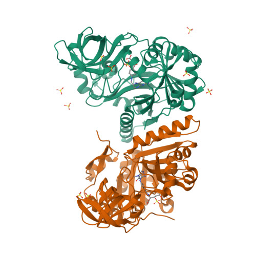
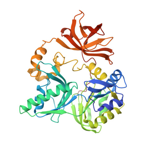
    T-protein, a component of the glycine cleavage system, catalyzes the formation of ammonia and 5,10-methylenetetrahydrofolate from the aminomethyl moiety of glycine attached to the lipoate cofactor of H-protein. Several mutations in the human T-protein gene cause non-ketotic hyperglycinemia. To gain insights into the effect of disease-causing mutations and the catalytic mechanism at the molecular level, crystal structures of human T-protein in free form and that bound to 5-methyltetrahydrofolate (5-CH3-H4folate) have been determined at 2.0 A and 2.6 A resolution, respectively. The overall structure consists of three domains arranged in a cloverleaf-like structure with the central cavity, where 5-CH3-H4folate is bound in a kinked shape with the pteridine group deeply buried into the hydrophobic pocket and the glutamyl group pointed to the C-terminal side surface. Most of the disease-related residues cluster around the cavity, forming extensive hydrogen bonding networks. These hydrogen bonding networks are employed in holding not only the folate-binding space but also the positions and the orientations of alpha-helix G and the following loop in the middle region, which seems to play a pivotal role in the T-protein catalysis. Structural and mutational analyses demonstrated that Arg292 interacts through water molecules with the folate polyglutamate tail, and that the invariant Asp101, located close to the N10 group of 5-CH3-H4folate, might play a key role in the initiation of the catalysis by increasing the nucleophilic character of the N10 atom of the folate substrate for the nucleophilic attack on the aminomethyl lipoate intermediate. A clever mechanism of recruiting the aminomethyl lipoate arm to the reaction site seems to function as a way of avoiding the release of toxic formaldehyde.


  • Organizational Affiliation

    Institute for Enzyme Research, The University of Tokushima, Tokushima 770-8503, Japan. ikeda@ier.tokushima-u.ac.jp


Macromolecules
Find similar proteins by:  (by identity cutoff)  |  3D Structure
Entity ID: 1
MoleculeChains Sequence LengthOrganismDetailsImage
Aminomethyltransferase
A, B
375Homo sapiensMutation(s): 0 
Gene Names: GCST
EC: 2.1.2.10
UniProt & NIH Common Fund Data Resources
Find proteins for P48728 (Homo sapiens)
Explore P48728 
Go to UniProtKB:  P48728
PHAROS:  P48728
GTEx:  ENSG00000145020 
Entity Groups  
Sequence Clusters30% Identity50% Identity70% Identity90% Identity95% Identity100% Identity
UniProt GroupP48728
Sequence Annotations
Expand
  • Reference Sequence
Small Molecules
Ligands 2 Unique
IDChains Name / Formula / InChI Key2D Diagram3D Interactions
THH
Query on THH

Download Ideal Coordinates CCD File 
K [auth A],
O [auth B]
N-[4-({[(6S)-2-AMINO-4-HYDROXY-5-METHYL-5,6,7,8-TETRAHYDROPTERIDIN-6-YL]METHYL}AMINO)BENZOYL]-L-GLUTAMIC ACID
C20 H25 N7 O6
ZNOVTXRBGFNYRX-STQMWFEESA-N
SO4
Query on SO4

Download Ideal Coordinates CCD File 
C [auth A]
D [auth A]
E [auth A]
F [auth A]
G [auth A]
SULFATE ION
O4 S
QAOWNCQODCNURD-UHFFFAOYSA-L
Experimental Data & Validation

Experimental Data

  • Method: X-RAY DIFFRACTION
  • Resolution: 2.60 Å
  • R-Value Free:  0.245 (Depositor), 0.170 (DCC) 
  • R-Value Work:  0.171 (Depositor), 0.170 (DCC) 
  • R-Value Observed: 0.171 (Depositor) 
Space Group: P 21 21 21
Unit Cell:
Length ( Å )Angle ( ˚ )
a = 54.67α = 90
b = 129.06β = 90
c = 135.76γ = 90
Software Package:
Software NamePurpose
CNSrefinement
DENZOdata reduction
SCALEPACKdata scaling
MOLREPphasing

Structure Validation

View Full Validation Report


Ligand Structure Quality Assessment 

Created with Raphaël 2.3.0Worse 01 BetterLigand structure goodness of fit to experimental dataBest fitted THHClick on this verticalbar to view details

Entry History 

Deposition Data

Revision History  (Full details and data files)

  • Version 1.0: 2005-08-16
    Type: Initial release
  • Version 1.1: 2008-04-30
    Changes: Version format compliance
  • Version 1.2: 2011-07-13
    Changes: Version format compliance
  • Version 1.3: 2023-10-25
    Changes: Data collection, Database references, Derived calculations, Refinement description