1TUK | pdb_00001tuk

Crystal structure of liganded type 2 non specific lipid transfer protein from wheat


Experimental Data Snapshot

  • Method: X-RAY DIFFRACTION
  • Resolution: 1.12 Å
  • R-Value Free: 
    0.162 (Depositor), 0.160 (DCC) 
  • R-Value Work: 
    0.137 (Depositor), 0.160 (DCC) 
  • R-Value Observed: 
    0.138 (Depositor) 

wwPDB Validation   3D Report Full Report


Ligand Structure Quality Assessment 

Created with Raphaël 2.3.0Worse 01 BetterLigand structure goodness of fit to experimental dataBest fitted PGMClick on this verticalbar to view details

This is version 1.5 of the entry. See complete history


Literature

Structure of a liganded type 2 non-specific lipid-transfer protein from wheat and the molecular basis of lipid binding.

Hoh, F.Pons, J.L.Gautier, M.F.de Lamotte, F.Dumas, C.

(2005) Acta Crystallogr D Biol Crystallogr 61: 397-406

  • DOI: https://doi.org/10.1107/S0907444905000417
  • Primary Citation of Related Structures:  
    1TUK

  • PubMed Abstract: 

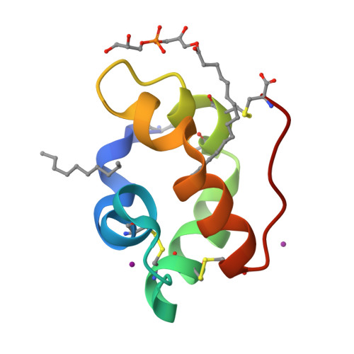
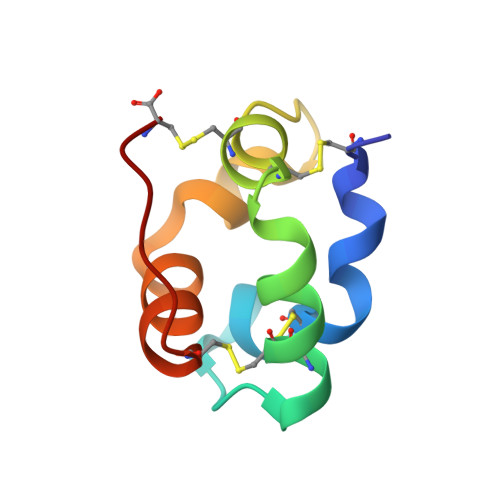
    In plants, a family of ubiquitous proteins named non-specific lipid-transfer proteins (ns-LTPs) facilitates the transfer of fatty acids, phospholipids and steroids between membranes. Recent data suggest that these secreted proteins play a key role in the formation of cuticular wax layers and in defence mechanisms against pathogens. In this study, X-ray crystallography has been used to examine the structural details of the interaction between a wheat type 2 ns-LTP and a lipid, L-alpha-palmitoyl-phosphatidyl glycerol. This crystal structure was solved ab initio at 1.12 A resolution by direct methods. The typical alpha-helical bundle fold of this protein is maintained by four disulfide bridges and delineates two hydrophobic cavities. The inner surface of the main cavity is lined by non-polar residues that provide a hydrophobic environment for the palmitoyl moiety of the lipid. The head-group region of this lipid protrudes from the surface and makes several polar interactions with a conserved patch of basic residues at the entrance of the pocket. The alkyl chain of a second lipid is bound within an adjacent smaller cavity. The structure shows that binding of the lipid tails to the protein involves extensive hydrophobic interactions.


  • Organizational Affiliation

    Centre de Biochimie Structurale, CNRS UMR5048-INSERM U554, 29 Rue de Navacelles, 34090 Montpellier, France.


Macromolecules
Find similar proteins by:  (by identity cutoff)  |  3D Structure
Entity ID: 1
MoleculeChains Sequence LengthOrganismDetailsImage
Nonspecific lipid-transfer protein 2G67Triticum aestivumMutation(s): 0 
UniProt
Find proteins for P82900 (Triticum aestivum)
Explore P82900 
Go to UniProtKB:  P82900
Entity Groups  
Sequence Clusters30% Identity50% Identity70% Identity90% Identity95% Identity100% Identity
UniProt GroupP82900
Sequence Annotations
Expand
  • Reference Sequence
Experimental Data & Validation

Experimental Data

  • Method: X-RAY DIFFRACTION
  • Resolution: 1.12 Å
  • R-Value Free:  0.162 (Depositor), 0.160 (DCC) 
  • R-Value Work:  0.137 (Depositor), 0.160 (DCC) 
  • R-Value Observed: 0.138 (Depositor) 
Space Group: C 1 2 1
Unit Cell:
Length ( Å )Angle ( ˚ )
a = 61.465α = 90
b = 29.32β = 127.65
c = 41.55γ = 90
Software Package:
Software NamePurpose
REFMACrefinement
MOSFLMdata reduction
CCP4data scaling
SnBphasing

Structure Validation

View Full Validation Report


Ligand Structure Quality Assessment 

Created with Raphaël 2.3.0Worse 01 BetterLigand structure goodness of fit to experimental dataBest fitted PGMClick on this verticalbar to view details

Entry History 

Deposition Data

Revision History  (Full details and data files)

  • Version 1.0: 2005-04-05
    Type: Initial release
  • Version 1.1: 2008-04-30
    Changes: Version format compliance
  • Version 1.2: 2011-07-13
    Changes: Version format compliance
  • Version 1.3: 2016-12-28
    Changes: Structure summary
  • Version 1.4: 2017-10-11
    Changes: Refinement description
  • Version 1.5: 2024-10-30
    Changes: Data collection, Database references, Derived calculations, Structure summary