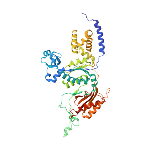
1T94

Crystal structure of the catalytic core of human DNA polymerase kappa


Experimental Data Snapshot

  • Method: X-RAY DIFFRACTION
  • Resolution: 2.40 Å
  • R-Value Free: 0.282 
  • R-Value Work: 0.246 
  • R-Value Observed: 0.246 

wwPDB Validation   3D Report Full Report


This is version 1.4 of the entry. See complete history


Literature

Crystal structure of the catalytic core of human DNA polymerase kappa.

Uljon, S.N.Johnson, R.E.Edwards, T.A.Prakash, S.Prakash, L.Aggarwal, A.K.

(2004) Structure 12: 1395-1404

  • DOI: https://doi.org/10.1016/j.str.2004.05.011
  • Primary Citation of Related Structures:  
    1T94

  • PubMed Abstract: 

    We present the crystal structure of the catalytic core of human DNA polymerase kappa (hPolkappa), the first structure of a human Y-family polymerase. hPolkappa is implicated in the proficient extension of mispaired primer termini on undamaged DNAs, and in the extension step of lesion bypass. The structure reveals a stubby "fingers" subdomain, which despite its small size appears to be tightly restrained with respect to a putative templating base. The structure also reveals a novel "thumb" subdomain that provides a basis for the importance of the N-terminal extension unique to hPolkappa. And, most surprisingly, the structure reveals the polymerase-associated domain (PAD) juxtaposed on the dorsal side of the "palm" subdomain, as opposed to the fingers subdomain. Together, these properties suggest that the hPolkappa active site is constrained at the site of the templating base and incoming nucleotide, but the polymerase is less constrained following translocation of the lesion.


  • Organizational Affiliation

    Structural Biology Program, Department of Physiology and Biophysics, Mount Sinai School of Medicine, Box 1677, 1425 Madison Avenue, New York, New York 10029, USA.


Macromolecules
Find similar proteins by:  (by identity cutoff)  |  3D Structure
Entity ID: 1
MoleculeChains Sequence LengthOrganismDetailsImage
polymerase (DNA directed) kappa
A, B
459Homo sapiensMutation(s): 0 
Gene Names: DINB
EC: 2.7.7.7
UniProt & NIH Common Fund Data Resources
Find proteins for Q9UBT6 (Homo sapiens)
Explore Q9UBT6 
Go to UniProtKB:  Q9UBT6
PHAROS:  Q9UBT6
GTEx:  ENSG00000122008 
Entity Groups  
Sequence Clusters30% Identity50% Identity70% Identity90% Identity95% Identity100% Identity
UniProt GroupQ9UBT6
Sequence Annotations
Expand
  • Reference Sequence
Experimental Data & Validation

Experimental Data

  • Method: X-RAY DIFFRACTION
  • Resolution: 2.40 Å
  • R-Value Free: 0.282 
  • R-Value Work: 0.246 
  • R-Value Observed: 0.246 
  • Space Group: P 21 21 21
Unit Cell:
Length ( Å )Angle ( ˚ )
a = 85.209α = 90
b = 109.46β = 90
c = 111.205γ = 90
Software Package:
Software NamePurpose
HKL-2000data collection
SCALEPACKdata scaling
SOLVEphasing
CNSrefinement
HKL-2000data reduction

Structure Validation

View Full Validation Report


Entry History 

Deposition Data

Revision History  (Full details and data files)

  • Version 1.0: 2004-08-31
    Type: Initial release
  • Version 1.1: 2008-04-30
    Changes: Version format compliance
  • Version 1.2: 2011-07-13
    Changes: Version format compliance
  • Version 1.3: 2018-01-31
    Changes: Experimental preparation
  • Version 1.4: 2024-02-14
    Changes: Data collection, Database references